 Movie
Movie Controller

[English] 日本語
 Yorodumi
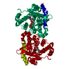
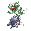
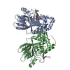
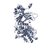
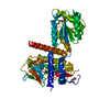
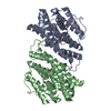
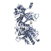
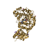
- PDB-1zct: structure of glycogenin truncated at residue 270 in a complex with UDP -
+ Open data

- Basic information
| Entry | Database: PDB / ID: 1zct | ||||||
|---|---|---|---|---|---|---|---|
| Title | structure of glycogenin truncated at residue 270 in a complex with UDP | ||||||
 Components Components | Glycogenin-1 | ||||||
 Keywords Keywords | TRANSFERASE / glycosyltransferase | ||||||
| Function / homology |  Function and homology information Function and homology information: / glycogenin glucosyltransferase / glycogenin glucosyltransferase activity / glycogen biosynthetic process / manganese ion binding / protein homodimerization activity / nucleus / cytoplasm Similarity search - Function | ||||||
| Biological species |  | ||||||
| Method |  X-RAY DIFFRACTION / SYNCHROTRON / MOLECULAR REPLACEMENT / Resolution: 2.6 Å X-RAY DIFFRACTION / SYNCHROTRON / MOLECULAR REPLACEMENT / Resolution: 2.6 Å | ||||||
 Authors Authors | Hurley, T.D. / Stout, S.L. / Miner, E. / Zhou, J. / Roach, P.J. | ||||||
 Citation Citation | Journal: J.Biol.Chem. / Year: 2005 Title: Requirements for catalysis in mammalian glycogenin. Authors: Hurley, T.D. / Stout, S. / Miner, E. / Zhou, J. / Roach, P.J. #1: Journal: J.Mol.Biol. / Year: 2002Title: Crystal structure of the autocatalytic initiator of glycogen biosynthesis, glycogenin Authors: Gibbons, B.J. / Roach, P.J. / Hurley, T.D. | ||||||
| History |
|
- Structure visualization
| Structure viewer | Molecule: MolmilJmol/JSmol |
|---|
- Downloads & links
-Download
| PDBx/mmCIF format | 1zct.cif.gz | 119.2 KB | Display | PDBx/mmCIF format |
|---|---|---|---|---|
| PDB format | pdb1zct.ent.gz | 90.5 KB | Display | PDB format |
| PDBx/mmJSON format | 1zct.json.gz | Tree view | PDBx/mmJSON format | |
| Others |  Other downloads Other downloads |
-Validation report
| Arichive directory | https://data.pdbj.org/pub/pdb/validation_reports/zc/1zctftp://data.pdbj.org/pub/pdb/validation_reports/zc/1zct | HTTPS FTP |
|---|
-Related structure data
| Related structure data | 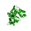 1zcuC  1zcvC 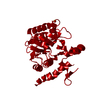 1zcyC 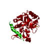 1zdfC 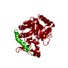 1zdgC  1ll3S S: Starting model for refinement C: citing same article ( |
|---|---|
| Similar structure data |


-Links
 PDBj
PDBj


- Assembly
| Deposited unit | 
| ||||||||
|---|---|---|---|---|---|---|---|---|---|
| 1 |
| ||||||||
| 2 | 
| ||||||||
| Unit cell |
|
-Components
| #1: Protein | Mass: 32504.529 Da / Num. of mol.: 2 Source method: isolated from a genetically manipulated source Source: (gene. exp.)  #2: Chemical |   Mass: 54.938 Da / Num. of mol.: 2 / Source method: obtained synthetically / Formula: Mn Mass: 54.938 Da / Num. of mol.: 2 / Source method: obtained synthetically / Formula: Mn#3: Chemical |   Type: RNA linking / Mass: 404.161 Da / Num. of mol.: 2 / Source method: obtained synthetically / Formula: C9H14N2O12P2 / Comment: UDP*YM Type: RNA linking / Mass: 404.161 Da / Num. of mol.: 2 / Source method: obtained synthetically / Formula: C9H14N2O12P2 / Comment: UDP*YM#4: Water | ChemComp-HOH / |  Mass: 18.015 Da / Num. of mol.: 55 / Source method: isolated from a natural source / Formula: H2O Mass: 18.015 Da / Num. of mol.: 55 / Source method: isolated from a natural source / Formula: H2O |
|---|
-Experimental details
-Experiment
| Experiment | Method: X-RAY DIFFRACTION / Number of used crystals: 1 |
|---|
- Sample preparation
| Crystal | Density Matthews: 3 Å3/Da / Density % sol: 57.4 % |
|---|---|
| Crystal grow | Temperature: 293 K / Method: vapor diffusion, hanging drop / pH: 4.6 Details: sodium acetate, PEG 4000, uridine-diphosphoglucose, manganese chloride, pH 4.6, VAPOR DIFFUSION, HANGING DROP, temperature 293K |
-Data collection
| Diffraction | Mean temperature: 93 K |
|---|---|
| Diffraction source | Source: SYNCHROTRON / Site: APS  / Beamline: 17-ID / Wavelength: 1 Å / Beamline: 17-ID / Wavelength: 1 Å |
| Detector | Type: ADSC QUANTUM 210 / Detector: CCD / Date: Nov 1, 2003 |
| Radiation | Monochromator: APS 17ID design / Protocol: SINGLE WAVELENGTH / Monochromatic (M) / Laue (L): M / Scattering type: x-ray |
| Radiation wavelength | Wavelength: 1 Å / Relative weight: 1 |
| Reflection | Resolution: 2.6→20 Å / Num. all: 22737 / Num. obs: 20872 / % possible obs: 91.2 % / Observed criterion σ(F): 0.2 / Observed criterion σ(I): 0.2 / Redundancy: 10.1 % / Biso Wilson estimate: 55.5 Å2 / Rmerge(I) obs: 0.063 / Χ2: 1.054 / Net I/σ(I): 28.2 |
| Reflection shell | Resolution: 2.6→2.69 Å / % possible obs: 53.8 % / Rmerge(I) obs: 0.208 / Mean I/σ(I) obs: 7.4 / Num. measured obs: 1224 / Χ2: 1.062 / % possible all: 53.8 |
-Phasing
| Phasing MR | Rfactor: 43.5 / Cor.coef. Fo:Fc: 60.1 / Cor.coef. Io to Ic: 54.6
|
|---|
- Processing
| Software |
| ||||||||||||||||||||||||||||
|---|---|---|---|---|---|---|---|---|---|---|---|---|---|---|---|---|---|---|---|---|---|---|---|---|---|---|---|---|---|
| Refinement | Method to determine structure: MOLECULAR REPLACEMENT Starting model: PDB ENTRY 1LL3 Resolution: 2.6→20 Å / Isotropic thermal model: restrained / Cross valid method: THROUGHOUT / σ(F): 0 / Stereochemistry target values: Engh & Huber
| ||||||||||||||||||||||||||||
| Solvent computation | Bsol: 31.617 Å2 | ||||||||||||||||||||||||||||
| Displacement parameters | Biso mean: 74.231 Å2
| ||||||||||||||||||||||||||||
| Refine analyze |
| ||||||||||||||||||||||||||||
| Refinement step | Cycle: LAST / Resolution: 2.6→20 Å
| ||||||||||||||||||||||||||||
| Refine LS restraints |
| ||||||||||||||||||||||||||||
| Xplor file |
|
