 Movie
Movie Controller


+ Open data

- Basic information
| Entry | Database: PDB / ID: 1ll3 | ||||||
|---|---|---|---|---|---|---|---|
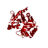
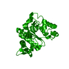
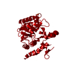
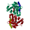
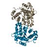
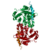
| Title | Crystal Structure of Rabbit Muscle Glycogenin | ||||||
 Components Components | GLYCOGENIN-1 | ||||||
 Keywords Keywords | TRANSFERASE / autocatalytic initiator of glycogen biosynthesis / glycogenin / retaining glycosyltransferase - family 8 / beta-alpha-beta Rossman-like nucleotide binding fold / DxD motif / non-proline cis peptide bond | ||||||
| Function / homology |  Function and homology information Function and homology information: / glycogenin glucosyltransferase / glycogenin glucosyltransferase activity / glycogen biosynthetic process / manganese ion binding / protein homodimerization activity / nucleus / cytoplasm Similarity search - Function | ||||||
| Biological species |  | ||||||
| Method |  X-RAY DIFFRACTION / SYNCHROTRON / MOLECULAR REPLACEMENT / Resolution: 1.9 Å X-RAY DIFFRACTION / SYNCHROTRON / MOLECULAR REPLACEMENT / Resolution: 1.9 Å | ||||||
 Authors Authors | Gibbons, B.J. / Roach, P.J. / Hurley, T.D. | ||||||
 Citation Citation | Journal: J.Mol.Biol. / Year: 2002 Title: Crystal structure of the autocatalytic initiator of glycogen biosynthesis, glycogenin. Authors: Gibbons, B.J. / Roach, P.J. / Hurley, T.D. | ||||||
| History |
|
- Structure visualization
| Structure viewer | Molecule: MolmilJmol/JSmol |
|---|
- Downloads & links
-Download
| PDBx/mmCIF format | 1ll3.cif.gz | 72 KB | Display | PDBx/mmCIF format |
|---|---|---|---|---|
| PDB format | pdb1ll3.ent.gz | 52.6 KB | Display | PDB format |
| PDBx/mmJSON format | 1ll3.json.gz | Tree view | PDBx/mmJSON format | |
| Others |  Other downloads Other downloads |
-Validation report
| Arichive directory | https://data.pdbj.org/pub/pdb/validation_reports/ll/1ll3ftp://data.pdbj.org/pub/pdb/validation_reports/ll/1ll3 | HTTPS FTP |
|---|
-Related structure data


-Links
 PDBj
PDBj

- Assembly
| Deposited unit | 
| |||||||||
|---|---|---|---|---|---|---|---|---|---|---|
| 1 | 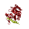
| |||||||||
| Unit cell |
| |||||||||
| Components on special symmetry positions |
| |||||||||
| Details | The asymetric unit contains one monomer. To construct the dimer apply the following symmetry operations: matrix:(-1.00000 -0.00001 0.00002)(0.00001 -1.00000 0.00002)(0.00002 0.00002 1.00000) translation:(-0.00052 -106.87773 0.00065) |
-Components
| #1: Protein | Mass: 37432.852 Da / Num. of mol.: 1 Source method: isolated from a genetically manipulated source Source: (gene. exp.)  | ||
|---|---|---|---|
| #2: Chemical |   Mass: 92.094 Da / Num. of mol.: 2 / Source method: obtained synthetically / Formula: C3H8O3 Mass: 92.094 Da / Num. of mol.: 2 / Source method: obtained synthetically / Formula: C3H8O3#3: Water | ChemComp-HOH / |  Mass: 18.015 Da / Num. of mol.: 245 / Source method: isolated from a natural source / Formula: H2O Mass: 18.015 Da / Num. of mol.: 245 / Source method: isolated from a natural source / Formula: H2O |
-Experimental details
-Experiment
| Experiment | Method: X-RAY DIFFRACTION / Number of used crystals: 1 |
|---|
- Sample preparation
| Crystal | Density Matthews: 2.5 Å3/Da / Density % sol: 50.72 % | ||||||||||||||||||||||||
|---|---|---|---|---|---|---|---|---|---|---|---|---|---|---|---|---|---|---|---|---|---|---|---|---|---|
| Crystal grow | Temperature: 298 K / Method: vapor diffusion, sitting drop / pH: 6.8 Details: 8-10 mg/mL glycogenin, 0.7 to 1.2 M ammonium sulphate, and 100 mM sodium phosphate buffer pH 6.6 to 6.9, pH 6.8, VAPOR DIFFUSION, SITTING DROP, temperature 298K | ||||||||||||||||||||||||
| Crystal grow | *PLUS Temperature: 4 ℃ / PH range low: 6.9 / PH range high: 6.6 | ||||||||||||||||||||||||
| Components of the solutions | *PLUS
|
-Data collection
| Diffraction | Mean temperature: 108 K |
|---|---|
| Diffraction source | Source: SYNCHROTRON / Site: NSLS  / Beamline: X12B / Wavelength: 1.079 / Beamline: X12B / Wavelength: 1.079 |
| Detector | Type: ADSC QUANTUM 4 / Detector: CCD / Date: Jun 4, 2001 |
| Radiation | Protocol: SINGLE WAVELENGTH / Monochromatic (M) / Laue (L): M / Scattering type: x-ray |
| Radiation wavelength | Wavelength: 1.079 Å / Relative weight: 1 |
| Reflection | Resolution: 1.9→30 Å / Num. all: 29958 / Num. obs: 29664 / % possible obs: 99 % / Observed criterion σ(F): 0 / Observed criterion σ(I): 0 / Rmerge(I) obs: 0.062 / Net I/σ(I): 23.1 |
| Reflection shell | Resolution: 1.9→1.97 Å / Rmerge(I) obs: 0.559 / Mean I/σ(I) obs: 2.4 / Num. unique all: 29807 / % possible all: 92.9 |
| Reflection | *PLUS Lowest resolution: 30 Å / Num. obs: 29807 / % possible obs: 98.4 % / Num. measured all: 162821 / Rmerge(I) obs: 0.062 |
| Reflection shell | *PLUS % possible obs: 92.9 % / Rmerge(I) obs: 0.559 |
- Processing
| Software |
| |||||||||||||||||||||||||||
|---|---|---|---|---|---|---|---|---|---|---|---|---|---|---|---|---|---|---|---|---|---|---|---|---|---|---|---|---|
| Refinement | Method to determine structure: MOLECULAR REPLACEMENT / Resolution: 1.9→30 Å / σ(F): 0 / Stereochemistry target values: Engh & Huber
| |||||||||||||||||||||||||||
| Displacement parameters | Biso mean: 36.8 Å2
| |||||||||||||||||||||||||||
| Refine analyze |
| |||||||||||||||||||||||||||
| Refinement step | Cycle: LAST / Resolution: 1.9→30 Å
| |||||||||||||||||||||||||||
| Refine LS restraints |
| |||||||||||||||||||||||||||
| Refinement | *PLUS Lowest resolution: 30 Å / Rfactor obs: 0.212 / Rfactor Rfree: 0.246 / Rfactor Rwork: 0.212 | |||||||||||||||||||||||||||
| Solvent computation | *PLUS | |||||||||||||||||||||||||||
| Displacement parameters | *PLUS | |||||||||||||||||||||||||||
| Refine LS restraints | *PLUS
|
