 Movie
Movie Controller


+ Open data

- Basic information
| Entry | Database: PDB / ID: 3usr | ||||||
|---|---|---|---|---|---|---|---|
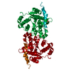
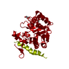
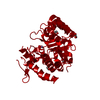
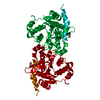
| Title | Structure of Y194F glycogenin mutant truncated at residue 270 | ||||||
 Components Components | Glycogenin-1 | ||||||
 Keywords Keywords | TRANSFERASE / Glucosyltransferase | ||||||
| Function / homology |  Function and homology information Function and homology information: / glycogenin glucosyltransferase / glycogenin glucosyltransferase activity / glycogen biosynthetic process / manganese ion binding / protein homodimerization activity / nucleus / cytoplasm Similarity search - Function | ||||||
| Biological species |  | ||||||
| Method |  X-RAY DIFFRACTION / SYNCHROTRON / FOURIER SYNTHESIS / Resolution: 2.1 Å X-RAY DIFFRACTION / SYNCHROTRON / FOURIER SYNTHESIS / Resolution: 2.1 Å | ||||||
 Authors Authors | Issoglio, F.M. / Carrizo, M.E. / Romero, J.M. / Curtino, J.A. | ||||||
 Citation Citation | Journal: J.Biol.Chem. / Year: 2012 Title: Mechanisms of monomeric and dimeric glycogenin autoglucosylation. Authors: Issoglio, F.M. / Carrizo, M.E. / Romero, J.M. / Curtino, J.A. | ||||||
| History |
|
- Structure visualization
| Structure viewer | Molecule: MolmilJmol/JSmol |
|---|
- Downloads & links
-Download
| PDBx/mmCIF format | 3usr.cif.gz | 68 KB | Display | PDBx/mmCIF format |
|---|---|---|---|---|
| PDB format | pdb3usr.ent.gz | 49.6 KB | Display | PDB format |
| PDBx/mmJSON format | 3usr.json.gz | Tree view | PDBx/mmJSON format | |
| Others |  Other downloads Other downloads |
-Validation report
| Arichive directory | https://data.pdbj.org/pub/pdb/validation_reports/us/3usrftp://data.pdbj.org/pub/pdb/validation_reports/us/3usr | HTTPS FTP |
|---|
-Related structure data


-Links
 PDBj
PDBj

- Assembly
| Deposited unit | 
| ||||||||
|---|---|---|---|---|---|---|---|---|---|
| 1 | 
| ||||||||
| 2 | 
| ||||||||
| Unit cell |
|
-Components
| #1: Protein | Mass: 32591.670 Da / Num. of mol.: 1 / Mutation: Y194F Source method: isolated from a genetically manipulated source Source: (gene. exp.)  | ||||
|---|---|---|---|---|---|
| #2: Chemical |   Mass: 92.094 Da / Num. of mol.: 2 / Source method: obtained synthetically / Formula: C3H8O3 Mass: 92.094 Da / Num. of mol.: 2 / Source method: obtained synthetically / Formula: C3H8O3#3: Chemical | ChemComp-CL / |   Mass: 35.453 Da / Num. of mol.: 1 / Source method: obtained synthetically / Formula: Cl Mass: 35.453 Da / Num. of mol.: 1 / Source method: obtained synthetically / Formula: Cl#4: Water | ChemComp-HOH / |  Mass: 18.015 Da / Num. of mol.: 119 / Source method: isolated from a natural source / Formula: H2O Mass: 18.015 Da / Num. of mol.: 119 / Source method: isolated from a natural source / Formula: H2O |
-Experimental details
-Experiment
| Experiment | Method: X-RAY DIFFRACTION / Number of used crystals: 1 |
|---|
- Sample preparation
| Crystal | Density Matthews: 2.76 Å3/Da / Density % sol: 55.5 % |
|---|---|
| Crystal grow | Temperature: 298 K / Method: vapor diffusion, hanging drop / pH: 6.5 Details: 12% PEGMME5000/0.2M ammonium sulfate/sodium MES , pH 6.5, VAPOR DIFFUSION, HANGING DROP, temperature 298K |
-Data collection
| Diffraction | Mean temperature: 100 K |
|---|---|
| Diffraction source | Source: SYNCHROTRON / Site: LNLS  / Beamline: D03B-MX1 / Wavelength: 1.43 Å / Beamline: D03B-MX1 / Wavelength: 1.43 Å |
| Detector | Type: MARRESEARCH / Detector: CCD / Date: Aug 18, 2010 / Details: mirrors |
| Radiation | Monochromator: Silicium curved crystal, with asymmetric 7.25 angle cut Protocol: SINGLE WAVELENGTH / Monochromatic (M) / Laue (L): M / Scattering type: x-ray |
| Radiation wavelength | Wavelength: 1.43 Å / Relative weight: 1 |
| Reflection | Resolution: 2.1→78.22 Å / Num. obs: 21489 / % possible obs: 99.9 % / Observed criterion σ(F): 0 / Observed criterion σ(I): 0 / Redundancy: 4 % / Rmerge(I) obs: 0.068 / Net I/σ(I): 8.8 |
| Reflection shell | Resolution: 2.1→2.21 Å / Redundancy: 4 % / Rmerge(I) obs: 0.295 / Mean I/σ(I) obs: 2.6 / Num. unique all: 3091 / % possible all: 99.9 |
- Processing
| Software |
| |||||||||||||||||||||||||||||||||||||||||||||||||||||||||||||||||
|---|---|---|---|---|---|---|---|---|---|---|---|---|---|---|---|---|---|---|---|---|---|---|---|---|---|---|---|---|---|---|---|---|---|---|---|---|---|---|---|---|---|---|---|---|---|---|---|---|---|---|---|---|---|---|---|---|---|---|---|---|---|---|---|---|---|---|
| Refinement | Method to determine structure: FOURIER SYNTHESIS / Resolution: 2.1→78.22 Å / Cor.coef. Fo:Fc: 0.943 / Cor.coef. Fo:Fc free: 0.916 / SU B: 3.766 / SU ML: 0.103 / Cross valid method: THROUGHOUT / σ(F): 0 / ESU R: 0.183 / ESU R Free: 0.168 / Stereochemistry target values: MAXIMUM LIKELIHOOD / Details: HYDROGENS HAVE BEEN ADDED IN THE RIDING POSITIONS
| |||||||||||||||||||||||||||||||||||||||||||||||||||||||||||||||||
| Solvent computation | Ion probe radii: 0.8 Å / Shrinkage radii: 0.8 Å / VDW probe radii: 1.4 Å / Solvent model: MASK | |||||||||||||||||||||||||||||||||||||||||||||||||||||||||||||||||
| Displacement parameters | Biso mean: 25.367 Å2
| |||||||||||||||||||||||||||||||||||||||||||||||||||||||||||||||||
| Refinement step | Cycle: LAST / Resolution: 2.1→78.22 Å
| |||||||||||||||||||||||||||||||||||||||||||||||||||||||||||||||||
| Refine LS restraints |
| |||||||||||||||||||||||||||||||||||||||||||||||||||||||||||||||||
| LS refinement shell | Resolution: 2.1→2.155 Å / Total num. of bins used: 20
|
