+ データを開く
- 基本情報
基本情報
| 登録情報 | データベース: PDB / ID: 1z9d | ||||||
|---|---|---|---|---|---|---|---|
| タイトル | Crystal structure of a putative uridylate kinase (UMP-kinase) from Streptococcus pyogenes | ||||||
 要素 要素 | uridylate kinase | ||||||
 キーワード キーワード | TRANSFERASE / Structural Genomics / Protein Structure Initiative / NYSGXRC / T1668 / pyrH / putative uridylate kinase / UMP-kinase / PSI / New York SGX Research Center for Structural Genomics | ||||||
| 機能・相同性 |  機能・相同性情報 機能・相同性情報UMP kinase / UMP kinase activity / 'de novo' CTP biosynthetic process / UDP biosynthetic process / ATP binding / cytoplasm 類似検索 - 分子機能 | ||||||
| 生物種 |  Streptococcus pyogenes (化膿レンサ球菌) Streptococcus pyogenes (化膿レンサ球菌) | ||||||
| 手法 |  X線回折 / X線回折 /  シンクロトロン / SAD aided by molecular replacement / 解像度: 2.8 Å シンクロトロン / SAD aided by molecular replacement / 解像度: 2.8 Å | ||||||
 データ登録者 データ登録者 | Rajashankar, K.R. / Kniewel, R. / Lee, K. / Lima, C.D. / Burley, S.K. / New York SGX Research Center for Structural Genomics (NYSGXRC) | ||||||
 引用 引用 |  ジャーナル: To be Published ジャーナル: To be Publishedタイトル: Crystal structure of a putative uridylate kinase (UMP-kinase) from Streptococcus pyogenes 著者: Rajashankar, K.R. / Kniewel, R. / Lee, K. / Lima, C.D. | ||||||
| 履歴 |
|
- 構造の表示
構造の表示
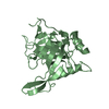
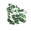
| 構造ビューア | 分子:  Molmil Molmil Jmol/JSmol Jmol/JSmol |
|---|
- ダウンロードとリンク
- ダウンロード
| PDBx/mmCIF形式 |  1z9d.cif.gz 1z9d.cif.gz | 146.1 KB | 表示 |  PDBx/mmCIF形式 PDBx/mmCIF形式 |
|---|---|---|---|---|
| PDB形式 |  pdb1z9d.ent.gz pdb1z9d.ent.gz | 116.1 KB | 表示 |  PDB形式 PDB形式 |
| PDBx/mmJSON形式 |  1z9d.json.gz 1z9d.json.gz | ツリー表示 |  PDBx/mmJSON形式 PDBx/mmJSON形式 | |
| その他 |  その他のダウンロード その他のダウンロード |
-検証レポート
| 文書・要旨 |  1z9d_validation.pdf.gz 1z9d_validation.pdf.gz | 464.1 KB | 表示 |  wwPDB検証レポート wwPDB検証レポート |
|---|---|---|---|---|
| 文書・詳細版 |  1z9d_full_validation.pdf.gz 1z9d_full_validation.pdf.gz | 477.4 KB | 表示 | |
| XML形式データ |  1z9d_validation.xml.gz 1z9d_validation.xml.gz | 27.9 KB | 表示 | |
| CIF形式データ |  1z9d_validation.cif.gz 1z9d_validation.cif.gz | 37.5 KB | 表示 | |
| アーカイブディレクトリ |  https://data.pdbj.org/pub/pdb/validation_reports/z9/1z9d https://data.pdbj.org/pub/pdb/validation_reports/z9/1z9d ftp://data.pdbj.org/pub/pdb/validation_reports/z9/1z9d ftp://data.pdbj.org/pub/pdb/validation_reports/z9/1z9d | HTTPS FTP |
-関連構造データ
| 類似構造データ | |
|---|---|
| その他のデータベース |
- リンク
リンク
- 集合体
集合体
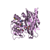
| 登録構造単位 | 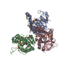
| ||||||||||
|---|---|---|---|---|---|---|---|---|---|---|---|
| 1 | 
| ||||||||||
| 2 | 
| ||||||||||
| 3 | 
| ||||||||||
| 4 | 
| ||||||||||
| 単位格子 |
| ||||||||||
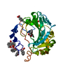
| 詳細 | UMP-Kinase from B. subtilis has a sequence identity of 40% to T1668 and is known to exist as a hexamer (a trimer of dimers). This fact has been experimentally tested via Gel filtration(C. Gagyi et. al. Eur. J. Biochem. 270, 3196-3204). However biologically active species are monomers. The hexamer can be generated by symmetry operation -X+1,-Y,Z. |
- 要素
要素
| #1: タンパク質 | 分子量: 27607.812 Da / 分子数: 3 / 由来タイプ: 組換発現 由来: (組換発現)  Streptococcus pyogenes (化膿レンサ球菌) Streptococcus pyogenes (化膿レンサ球菌)遺伝子: pyrH / プラスミド: PET T7 / 発現宿主:  参照: UniProt: P65938, 転移酵素; リンを含む基を移すもの; リン酸基に移すもの #2: 化合物 | ChemComp-SO4 / #3: 水 | ChemComp-HOH / | Has protein modification | Y | |
|---|
-実験情報
-実験
| 実験 | 手法:  X線回折 / 使用した結晶の数: 1 X線回折 / 使用した結晶の数: 1 |
|---|
- 試料調製
試料調製
| 結晶 | マシュー密度: 2.68 Å3/Da / 溶媒含有率: 53.72 % |
|---|---|
| 結晶化 | 温度: 291 K / 手法: 蒸気拡散法, ハンギングドロップ法 / pH: 8.5 詳細: 2M Ammonium sulfate, 0.15M Tris pH 8.5, VAPOR DIFFUSION, HANGING DROP, temperature 291K |
-データ収集
| 回折 | 平均測定温度: 100 K |
|---|---|
| 放射光源 | 由来:  シンクロトロン / サイト: シンクロトロン / サイト:  APS APS  / ビームライン: 31-ID / 波長: 0.98 Å / ビームライン: 31-ID / 波長: 0.98 Å |
| 検出器 | タイプ: MARRESEARCH / 検出器: CCD / 日付: 2004年6月26日 / 詳細: Diamond monochromator and downstream mirror |
| 放射 | モノクロメーター: Flat Diamond 111 / プロトコル: SINGLE WAVELENGTH / 単色(M)・ラウエ(L): M / 散乱光タイプ: x-ray |
| 放射波長 | 波長: 0.98 Å / 相対比: 1 |
| 反射 | 解像度: 2.8→20 Å / Num. all: 38306 / Num. obs: 38306 / % possible obs: 93.1 % / Observed criterion σ(I): -3 / 冗長度: 11.04 % / Biso Wilson estimate: 59.5 Å2 / Rsym value: 0.08 |
| 反射 シェル | 解像度: 2.8→2.9 Å / Mean I/σ(I) obs: 2.05 / Num. unique all: 3849 / Rsym value: 0.382 / % possible all: 92.9 |
- 解析
解析
| ソフトウェア |
| ||||||||||||||||||||||||||||||||||||
|---|---|---|---|---|---|---|---|---|---|---|---|---|---|---|---|---|---|---|---|---|---|---|---|---|---|---|---|---|---|---|---|---|---|---|---|---|---|
| 精密化 | 構造決定の手法: SAD aided by molecular replacement / 解像度: 2.8→19.72 Å / Rfactor Rfree error: 0.006 / Data cutoff high absF: 222033.22 / Data cutoff low absF: 0 / Isotropic thermal model: RESTRAINED / 交差検証法: THROUGHOUT / σ(F): 0 / 立体化学のターゲット値: Engh & Huber 詳細: A molecular replacement solution was obtained using a model derived from pdb entry 1YBD. MR phases were used to locate Se sites. Experimental phases were calculated using a Se-substructure ...詳細: A molecular replacement solution was obtained using a model derived from pdb entry 1YBD. MR phases were used to locate Se sites. Experimental phases were calculated using a Se-substructure containing 26 Se sites. Nine sulfate groups were located. Sulfates D4 - D9 mimic phosphate group of ATP at the ATP binding pocket.
| ||||||||||||||||||||||||||||||||||||
| 溶媒の処理 | 溶媒モデル: FLAT MODEL / Bsol: 11.7658 Å2 / ksol: 0.318634 e/Å3 | ||||||||||||||||||||||||||||||||||||
| 原子変位パラメータ | Biso mean: 41.5 Å2
| ||||||||||||||||||||||||||||||||||||
| Refine analyze |
| ||||||||||||||||||||||||||||||||||||
| 精密化ステップ | サイクル: LAST / 解像度: 2.8→19.72 Å
| ||||||||||||||||||||||||||||||||||||
| 拘束条件 |
| ||||||||||||||||||||||||||||||||||||
| LS精密化 シェル | 解像度: 2.8→2.97 Å / Rfactor Rfree error: 0.024 / Total num. of bins used: 6
| ||||||||||||||||||||||||||||||||||||
| Xplor file |
|
 ムービー
ムービー コントローラー





 PDBj
PDBj


