 Movie
Movie Controller

[English] 日本語
 Yorodumi
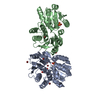
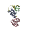
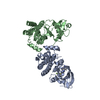
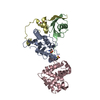
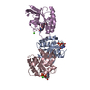
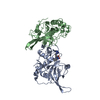
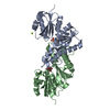
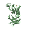
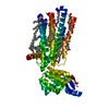
- PDB-1z5s: Crystal structure of a complex between UBC9, SUMO-1, RANGAP1 and ... -
+ Open data

- Basic information
| Entry | Database: PDB / ID: 1z5s | ||||||
|---|---|---|---|---|---|---|---|
| Title | Crystal structure of a complex between UBC9, SUMO-1, RANGAP1 and NUP358/RANBP2 | ||||||
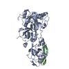
 Components Components |
| ||||||
 Keywords Keywords | LIGASE / E3 / SUMO / UBC9 / NUCLEAR PORE COMPLEX | ||||||
| Function / homology |  Function and homology information Function and homology informationcellular response to vasopressin / cytoplasmic periphery of the nuclear pore complex / SUMO conjugating enzyme activity / SUMO ligase complex / SUMO ligase activity / protein localization to nuclear pore / RING-like zinc finger domain binding / SUMOylation of nuclear envelope proteins / negative regulation of transcription initiation by RNA polymerase II / annulate lamellae ...cellular response to vasopressin / cytoplasmic periphery of the nuclear pore complex / SUMO conjugating enzyme activity / SUMO ligase complex / SUMO ligase activity / protein localization to nuclear pore / RING-like zinc finger domain binding / SUMOylation of nuclear envelope proteins / negative regulation of transcription initiation by RNA polymerase II / annulate lamellae / transferase complex / HLH domain binding / SUMO is proteolytically processed / Negative regulation of activity of TFAP2 (AP-2) family transcription factors / SUMO is conjugated to E1 (UBA2:SAE1) / SUMO is transferred from E1 to E2 (UBE2I, UBC9) / Vitamin D (calciferol) metabolism / negative regulation of action potential / nuclear pore cytoplasmic filaments / nuclear stress granule / PML body organization / mitotic nuclear membrane reassembly / Nuclear Pore Complex (NPC) Disassembly / small protein activating enzyme binding / synaptonemal complex / Regulation of Glucokinase by Glucokinase Regulatory Protein / Defective TPR may confer susceptibility towards thyroid papillary carcinoma (TPC) / activation of GTPase activity / nuclear inclusion body / Transport of Ribonucleoproteins into the Host Nucleus / nuclear pore nuclear basket / Transport of the SLBP independent Mature mRNA / Transport of the SLBP Dependant Mature mRNA / SUMOylation of immune response proteins / NS1 Mediated Effects on Host Pathways / SUMOylation of SUMOylation proteins / regulation of calcium ion transmembrane transport / Transport of Mature mRNA Derived from an Intronless Transcript / negative regulation of protein export from nucleus / SUMOylation of DNA methylation proteins / Maturation of nucleoprotein / Rev-mediated nuclear export of HIV RNA / nuclear export / Nuclear import of Rev protein / XY body / SUMOylation of RNA binding proteins / NEP/NS2 Interacts with the Cellular Export Machinery / Transferases; Acyltransferases; Aminoacyltransferases / regulation of cardiac muscle cell contraction / SUMO transferase activity / Transport of Mature mRNA derived from an Intron-Containing Transcript / tRNA processing in the nucleus / Postmitotic nuclear pore complex (NPC) reformation / kinase activator activity / aggresome / centrosome localization / Maturation of nucleoprotein / regulation of gluconeogenesis / Viral Messenger RNA Synthesis / nucleocytoplasmic transport / NLS-bearing protein import into nucleus / negative regulation of protein import into nucleus / SUMOylation of ubiquitinylation proteins / Vpr-mediated nuclear import of PICs / transcription factor binding / ubiquitin-specific protease binding / cellular response to cadmium ion / SUMOylation of transcription factors / roof of mouth development / SUMOylation of DNA replication proteins / ubiquitin-like protein ligase binding / Regulation of HSF1-mediated heat shock response / protein sumoylation / potassium channel regulator activity / Regulation of IFNG signaling / nuclear pore / transporter activator activity / mRNA transport / postsynaptic cytosol / response to axon injury / SUMOylation of DNA damage response and repair proteins / SARS-CoV-1 targets host intracellular signalling and regulatory pathways / presynaptic cytosol / axon cytoplasm / Amplification of signal from unattached kinetochores via a MAD2 inhibitory signal / Transcriptional and post-translational regulation of MITF-M expression and activity / SUMOylation of transcription cofactors / Mitotic Prometaphase / EML4 and NUDC in mitotic spindle formation / intracellular glucose homeostasis / Meiotic synapsis / Resolution of Sister Chromatid Cohesion / SUMOylation of chromatin organization proteins / protein modification process / transcription coregulator binding / GTPase activator activity / HCMV Late Events / response to amphetamine / Regulation of endogenous retroelements by KRAB-ZFP proteins / SUMOylation of intracellular receptors Similarity search - Function | ||||||
| Biological species |  Homo sapiens (human) Homo sapiens (human) | ||||||
| Method |  X-RAY DIFFRACTION / SYNCHROTRON / MOLECULAR REPLACEMENT / Resolution: 3.01 Å X-RAY DIFFRACTION / SYNCHROTRON / MOLECULAR REPLACEMENT / Resolution: 3.01 Å | ||||||
 Authors Authors | Reverter, D. / Lima, C.D. | ||||||
 Citation Citation | Journal: Nature / Year: 2005 Title: Insights into E3 ligase activity revealed by a SUMO-RanGAP1-Ubc9-Nup358 complex. Authors: Reverter, D. / Lima, C.D. | ||||||
| History |
| ||||||
| Remark 999 | SEQUENCE COVALENT ISOPEPTIDE BOND BETWEEN RANGAP1 LYS524 AND SUMO C-TERMINUS GLY97. |
- Structure visualization
| Structure viewer | Molecule: MolmilJmol/JSmol |
|---|
- Downloads & links
-Download
| PDBx/mmCIF format | 1z5s.cif.gz | 104.4 KB | Display | PDBx/mmCIF format |
|---|---|---|---|---|
| PDB format | pdb1z5s.ent.gz | 78.8 KB | Display | PDB format |
| PDBx/mmJSON format | 1z5s.json.gz | Tree view | PDBx/mmJSON format | |
| Others |  Other downloads Other downloads |
-Validation report
| Arichive directory | https://data.pdbj.org/pub/pdb/validation_reports/z5/1z5sftp://data.pdbj.org/pub/pdb/validation_reports/z5/1z5s | HTTPS FTP |
|---|
-Related structure data
| Related structure data | |
|---|---|
| Similar structure data |

-Links
 PDBj
PDBj


- Assembly
| Deposited unit | 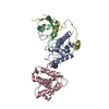
| ||||||||
|---|---|---|---|---|---|---|---|---|---|
| 1 |
| ||||||||
| Unit cell |
|
-Components
| #1: Protein | Mass: 18030.814 Da / Num. of mol.: 1 Source method: isolated from a genetically manipulated source Source: (gene. exp.) Homo sapiens (human) / Gene: UBE2I, UBC9, UBCE9 / Plasmid: PET28B / Species (production host): Escherichia coli / Production host:  |
|---|---|
| #2: Protein | Mass: 9473.775 Da / Num. of mol.: 1 Source method: isolated from a genetically manipulated source Source: (gene. exp.) Homo sapiens (human) / Gene: UBL1, SMT3C, SMT3H3 / Plasmid: PET28B / Species (production host): Escherichia coli / Production host: |
| #3: Protein | Mass: 18686.607 Da / Num. of mol.: 1 / Fragment: C-terminal domain Source method: isolated from a genetically manipulated source Source: (gene. exp.) Homo sapiens (human) / Gene: RANGAP1 / Plasmid: PSMT3 / Species (production host): Escherichia coli / Production host: |
| #4: Protein | Mass: 9544.638 Da / Num. of mol.: 1 / Fragment: IR1-M domain Source method: isolated from a genetically manipulated source Source: (gene. exp.) Homo sapiens (human) / Gene: RANBP2, NUP358 / Plasmid: PSMT3 / Species (production host): Escherichia coli / Production host: |
| #5: Water | ChemComp-HOH /  Mass: 18.015 Da / Num. of mol.: 28 / Source method: isolated from a natural source / Formula: H2O Mass: 18.015 Da / Num. of mol.: 28 / Source method: isolated from a natural source / Formula: H2O |
| Has protein modification | Y |
-Experimental details
-Experiment
| Experiment | Method: X-RAY DIFFRACTION / Number of used crystals: 1 |
|---|
- Sample preparation
| Crystal | Density Matthews: 3.81 Å3/Da / Density % sol: 67.73 % |
|---|---|
| Crystal grow | Temperature: 291 K / Method: vapor diffusion, hanging drop / pH: 5 Details: 18% PEG4000 (w/v), 0.1 M sodium citrate, 0.2 M ammonium acetate, pH 5.0, VAPOR DIFFUSION, HANGING DROP, temperature 291K |
-Data collection
| Diffraction | Mean temperature: 100 K |
|---|---|
| Diffraction source | Source: SYNCHROTRON / Site: APS  / Beamline: 31-ID / Wavelength: 0.9793 Å / Beamline: 31-ID / Wavelength: 0.9793 Å |
| Detector | Type: MARRESEARCH / Detector: CCD / Date: Aug 24, 2004 |
| Radiation | Monochromator: diamond / Protocol: SINGLE WAVELENGTH / Monochromatic (M) / Laue (L): M / Scattering type: x-ray |
| Radiation wavelength | Wavelength: 0.9793 Å / Relative weight: 1 |
| Reflection | Resolution: 3→50 Å / Num. obs: 16464 / % possible obs: 97 % / Observed criterion σ(F): 0 / Observed criterion σ(I): 0 / Biso Wilson estimate: 74 Å2 / Rmerge(I) obs: 0.075 / Net I/σ(I): 15.1 |
| Reflection shell | Resolution: 3→3.11 Å / Rmerge(I) obs: 0.464 / Mean I/σ(I) obs: 2.1 / % possible all: 91.8 |
- Processing
| Software |
| ||||||||||||||||||||||||||||||||||||
|---|---|---|---|---|---|---|---|---|---|---|---|---|---|---|---|---|---|---|---|---|---|---|---|---|---|---|---|---|---|---|---|---|---|---|---|---|---|
| Refinement | Method to determine structure: MOLECULAR REPLACEMENT Starting model: RANGAP1-UBC9 COMPLEX Resolution: 3.01→29.69 Å / Rfactor Rfree error: 0.01 / Data cutoff high absF: 2281669.87 / Data cutoff low absF: 0 / Isotropic thermal model: RESTRAINED / Cross valid method: THROUGHOUT / σ(F): 0 / Stereochemistry target values: Engh & Huber / Details: BULK SOLVENT MODEL USED
| ||||||||||||||||||||||||||||||||||||
| Solvent computation | Solvent model: FLAT MODEL / Bsol: 13.936 Å2 / ksol: 0.267286 e/Å3 | ||||||||||||||||||||||||||||||||||||
| Displacement parameters | Biso mean: 90 Å2
| ||||||||||||||||||||||||||||||||||||
| Refine analyze |
| ||||||||||||||||||||||||||||||||||||
| Refinement step | Cycle: LAST / Resolution: 3.01→29.69 Å
| ||||||||||||||||||||||||||||||||||||
| Refine LS restraints |
| ||||||||||||||||||||||||||||||||||||
| LS refinement shell | Resolution: 3.01→3.19 Å / Rfactor Rfree error: 0.039 / Total num. of bins used: 6
| ||||||||||||||||||||||||||||||||||||
| Xplor file |
|
