 Movie
Movie Controller

[English] 日本語
 Yorodumi
- PDB-1z3v: Structure of Phanerochaete chrysosporium cellobiohydrolase Cel7D ... -
+ Open data

- Basic information
| Entry | Database: PDB / ID: 1z3v | ||||||||||||
|---|---|---|---|---|---|---|---|---|---|---|---|---|---|
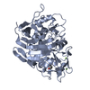
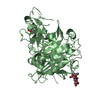
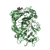
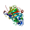
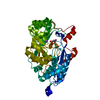
| Title | Structure of Phanerochaete chrysosporium cellobiohydrolase Cel7D (CBH58) in complex with lactose | ||||||||||||
 Components Components | cellulase | ||||||||||||
 Keywords Keywords | HYDROLASE / Beta sandwich | ||||||||||||
| Function / homology |  Function and homology information Function and homology informationHydrolases; Glycosylases; Glycosidases, i.e. enzymes that hydrolyse O- and S-glycosyl compounds / cellulose binding / cellulose catabolic process / hydrolase activity, hydrolyzing O-glycosyl compounds / extracellular region Similarity search - Function | ||||||||||||
| Biological species |  Phanerochaete chrysosporium (fungus) Phanerochaete chrysosporium (fungus) | ||||||||||||
| Method |  X-RAY DIFFRACTION / SYNCHROTRON / MOLECULAR REPLACEMENT / Resolution: 1.61 Å X-RAY DIFFRACTION / SYNCHROTRON / MOLECULAR REPLACEMENT / Resolution: 1.61 Å | ||||||||||||
 Authors Authors | Ubhayasekera, W. / Munoz, I.G. / Stahlberg, J. / Mowbray, S.L. | ||||||||||||
 Citation Citation | Journal: Febs J. / Year: 2005 Title: Structures of Phanerochaete chrysosporium Cel7D in complex with product and inhibitors Authors: Ubhayasekera, W. / Munoz, I.G. / Vasella, A. / Stahlberg, J. / Mowbray, S.L. #1: Journal: J.Mol.Biol. / Year: 2001Title: Family 7 cellobiohydrolases from Phanerochaete chrysosporium: crystal structure of the catalytic module of Cel7D (CBH58) at 1.32 A resolution and homology models of the isozymes Authors: Munoz, I.G. / Ubhayasekera, W. / Henriksson, H. / Szabo, I. / Pettersson, G. / Johansson, G. / Mowbray, S.L. / Stahlberg, J. #2: Journal: ACTA CRYSTALLOGR.,SECT.D / Year: 2003Title: The catalytic module of Cel7D from Phanerochaete chrysosporium as a chiral selector: structural studies of its complex with the beta blocker (R)-propranolol Authors: Munoz, I.G. / Mowbray, S.L. / Stahlberg, J. | ||||||||||||
| History |
|
- Structure visualization
| Structure viewer | Molecule: MolmilJmol/JSmol |
|---|
- Downloads & links
-Download
| PDBx/mmCIF format | 1z3v.cif.gz | 184.6 KB | Display | PDBx/mmCIF format |
|---|---|---|---|---|
| PDB format | pdb1z3v.ent.gz | 145.2 KB | Display | PDB format |
| PDBx/mmJSON format | 1z3v.json.gz | Tree view | PDBx/mmJSON format | |
| Others |  Other downloads Other downloads |
-Validation report
| Summary document | 1z3v_validation.pdf.gz | 800.4 KB | Display | wwPDB validaton report |
|---|---|---|---|---|
| Full document | 1z3v_full_validation.pdf.gz | 804.7 KB | Display | |
| Data in XML | 1z3v_validation.xml.gz | 20.6 KB | Display | |
| Data in CIF | 1z3v_validation.cif.gz | 30 KB | Display | |
| Arichive directory | https://data.pdbj.org/pub/pdb/validation_reports/z3/1z3vftp://data.pdbj.org/pub/pdb/validation_reports/z3/1z3v | HTTPS FTP |
-Related structure data
| Related structure data |  1z3tC  1z3wC  1gpiS S: Starting model for refinement C: citing same article ( |
|---|---|
| Similar structure data |


-Links
 PDBj
PDBj


- Assembly
| Deposited unit | 
| ||||||||
|---|---|---|---|---|---|---|---|---|---|
| 1 |
| ||||||||
| Unit cell |
|
-Components
| #1: Protein | Mass: 45777.141 Da / Num. of mol.: 1 / Fragment: Catalytic module / Source method: isolated from a natural source / Source: (natural) Phanerochaete chrysosporium (fungus) / Strain: K3References: UniProt: Q7LIJ0, cellulose 1,4-beta-cellobiosidase (non-reducing end) |
|---|---|
| #2: Polysaccharide | beta-D-galactopyranose-(1-4)-beta-D-glucopyranose / beta-lactose  Source method: isolated from a genetically manipulated source Details: oligosaccharide / References: beta-lactose |
| #3: Sugar | ChemComp-NAG /   Type: D-saccharide, beta linking / Mass: 221.208 Da / Num. of mol.: 1 Type: D-saccharide, beta linking / Mass: 221.208 Da / Num. of mol.: 1Source method: isolated from a genetically manipulated source Formula: C8H15NO6 |
| #4: Water | ChemComp-HOH /  Mass: 18.015 Da / Num. of mol.: 271 / Source method: isolated from a natural source / Formula: H2O Mass: 18.015 Da / Num. of mol.: 271 / Source method: isolated from a natural source / Formula: H2O |
| Has protein modification | Y |
-Experimental details
-Experiment
| Experiment | Method: X-RAY DIFFRACTION / Number of used crystals: 1 |
|---|
- Sample preparation
| Crystal | Density Matthews: 2.1 Å3/Da / Density % sol: 42.2 % |
|---|---|
| Crystal grow | Temperature: 294 K / Method: vapor diffusion, hanging drop / pH: 7 Details: TRIS-HCl, calcium chloride, PEG 5000, glycerol, pH 7.0, VAPOR DIFFUSION, HANGING DROP, temperature 294K |
-Data collection
| Diffraction | Mean temperature: 100 K |
|---|---|
| Diffraction source | Source: SYNCHROTRON / Site: ESRF  / Beamline: ID14-4 / Wavelength: 0.9322 Å / Beamline: ID14-4 / Wavelength: 0.9322 Å |
| Detector | Type: ADSC QUANTUM 4 / Detector: CCD / Date: Dec 11, 1999 |
| Radiation | Monochromator: Diamond / Protocol: SINGLE WAVELENGTH / Monochromatic (M) / Laue (L): M / Scattering type: x-ray |
| Radiation wavelength | Wavelength: 0.9322 Å / Relative weight: 1 |
| Reflection | Resolution: 1.6→50 Å / Num. all: 50272 / Num. obs: 50272 / % possible obs: 99.3 % / Observed criterion σ(F): 0 / Observed criterion σ(I): 0 / Redundancy: 3.4 % / Rmerge(I) obs: 0.072 / Net I/σ(I): 14.8 |
| Reflection shell | Resolution: 1.6→1.63 Å / Rmerge(I) obs: 0.238 / Mean I/σ(I) obs: 8.1 / Num. unique all: 3286 / % possible all: 99.6 |
- Processing
| Software |
| ||||||||||||||||||||||||||||||||||||||||||||||||||||||||||||||||||||||||||||||||||||||||||
|---|---|---|---|---|---|---|---|---|---|---|---|---|---|---|---|---|---|---|---|---|---|---|---|---|---|---|---|---|---|---|---|---|---|---|---|---|---|---|---|---|---|---|---|---|---|---|---|---|---|---|---|---|---|---|---|---|---|---|---|---|---|---|---|---|---|---|---|---|---|---|---|---|---|---|---|---|---|---|---|---|---|---|---|---|---|---|---|---|---|---|---|
| Refinement | Method to determine structure: MOLECULAR REPLACEMENT Starting model: PDB entry 1GPI Resolution: 1.61→39.22 Å / Cor.coef. Fo:Fc: 0.966 / Cor.coef. Fo:Fc free: 0.954 / SU B: 4.213 / SU ML: 0.062 / Cross valid method: THROUGHOUT / σ(F): 0 / σ(I): 0 / ESU R: 0.123 / ESU R Free: 0.09 / Stereochemistry target values: Engh & Huber
| ||||||||||||||||||||||||||||||||||||||||||||||||||||||||||||||||||||||||||||||||||||||||||
| Solvent computation | Ion probe radii: 0.8 Å / Shrinkage radii: 0.8 Å / VDW probe radii: 1.4 Å / Solvent model: BABINET MODEL WITH MASK | ||||||||||||||||||||||||||||||||||||||||||||||||||||||||||||||||||||||||||||||||||||||||||
| Displacement parameters | Biso mean: 22.962 Å2
| ||||||||||||||||||||||||||||||||||||||||||||||||||||||||||||||||||||||||||||||||||||||||||
| Refinement step | Cycle: LAST / Resolution: 1.61→39.22 Å
| ||||||||||||||||||||||||||||||||||||||||||||||||||||||||||||||||||||||||||||||||||||||||||
| Refine LS restraints |
| ||||||||||||||||||||||||||||||||||||||||||||||||||||||||||||||||||||||||||||||||||||||||||
| LS refinement shell | Resolution: 1.606→1.648 Å / Total num. of bins used: 20
|
