 Movie
Movie Controller

[English] 日本語
 Yorodumi
- PDB-1gpi: Cellobiohydrolase Cel7D (CBH 58) from Phanerochaete chrysosporium... -
+ Open data

- Basic information
| Entry | Database: PDB / ID: 1gpi | |||||||||
|---|---|---|---|---|---|---|---|---|---|---|
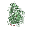
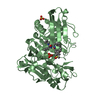
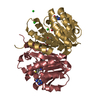
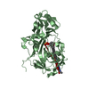
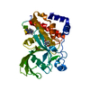
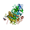
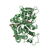
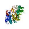
| Title | Cellobiohydrolase Cel7D (CBH 58) from Phanerochaete chrysosporium. Catalytic module at 1.32 Angstrom resolution | |||||||||
 Components Components | EXOGLUCANASE I | |||||||||
 Keywords Keywords | HYDROLASE / GLYCOSIDASE / CELLULASE / BETA-GLUCANASE / GLYCOPROTEIN / CELLULOSE DEGRADATION / ENZYME / REACTION CENTER / EXTRACELLULAR / EXOGLUCANASE | |||||||||
| Function / homology |  Function and homology information Function and homology informationcellulose binding / Hydrolases; Glycosylases; Glycosidases, i.e. enzymes that hydrolyse O- and S-glycosyl compounds / cellulase activity / cellulose catabolic process / extracellular region Similarity search - Function | |||||||||
| Biological species |  PHANEROCHAETE CHRYSOSPORIUM (fungus) PHANEROCHAETE CHRYSOSPORIUM (fungus) | |||||||||
| Method |  X-RAY DIFFRACTION / SYNCHROTRON / MOLECULAR REPLACEMENT / Resolution: 1.32 Å X-RAY DIFFRACTION / SYNCHROTRON / MOLECULAR REPLACEMENT / Resolution: 1.32 Å | |||||||||
 Authors Authors | Munoz, I.G. / Mowbray, S.L. / Stahlberg, J. | |||||||||
 Citation Citation | Journal: J.Mol.Biol. / Year: 2001 Title: Family 7 Cellobiohydrolases from Phanerochaete Chrysosporium: Crystal Structure of the Catalytic Module of Cel7D (Cbh58) at 1.32 Angstrom Resolution and Homology Models of the Isozymes. Authors: Munoz, I.G. / Ubhayasekera, W. / Henriksson, H. / Szabo, I. / Pettersson, G. / Johansson, G. / Mowbray, S.L. / Stahlberg, J. | |||||||||
| History |
| |||||||||
| Remark 700 | SHEET THE SHEET STRUCTURE OF THIS MOLECULE IS BIFURCATED. IN ORDER TO REPRESENT THIS FEATURE IN ... SHEET THE SHEET STRUCTURE OF THIS MOLECULE IS BIFURCATED. IN ORDER TO REPRESENT THIS FEATURE IN THE SHEET RECORDS BELOW, TWO SHEETS ARE DEFINED. |
- Structure visualization
| Structure viewer | Molecule: MolmilJmol/JSmol |
|---|
- Downloads & links
-Download
| PDBx/mmCIF format | 1gpi.cif.gz | 102 KB | Display | PDBx/mmCIF format |
|---|---|---|---|---|
| PDB format | pdb1gpi.ent.gz | 77.8 KB | Display | PDB format |
| PDBx/mmJSON format | 1gpi.json.gz | Tree view | PDBx/mmJSON format | |
| Others |  Other downloads Other downloads |
-Validation report
| Arichive directory | https://data.pdbj.org/pub/pdb/validation_reports/gp/1gpiftp://data.pdbj.org/pub/pdb/validation_reports/gp/1gpi | HTTPS FTP |
|---|
-Related structure data
| Related structure data | 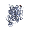 2celS S: Starting model for refinement |
|---|---|
| Similar structure data |


-Links
 PDBj
PDBj- Assembly
| Deposited unit | 
| ||||||||
|---|---|---|---|---|---|---|---|---|---|
| 1 |
| ||||||||
| Unit cell |
| ||||||||
| Components on special symmetry positions |
|
-Components
| #1: Protein | Mass: 45777.141 Da / Num. of mol.: 1 / Fragment: CATALYTIC MODULE, RESIDUES 19-450 / Source method: isolated from a natural source Details: EXTRACELLULAR PROTEIN OBTAINED FROM THE FED-BATCH CULTIVATION OF P. CHRYSOSPORIUM STRAIN K3 USING CELLULOSE (AVICEL) AS A CARBON SOURCE Source: (natural) PHANEROCHAETE CHRYSOSPORIUM (fungus) / Strain: K3References: UniProt: Q09431, UniProt: Q7LIJ0*PLUS, cellulose 1,4-beta-cellobiosidase (non-reducing end) | ||
|---|---|---|---|
| #2: Sugar | ChemComp-NAG /   Type: D-saccharide, beta linking / Mass: 221.208 Da / Num. of mol.: 1 Type: D-saccharide, beta linking / Mass: 221.208 Da / Num. of mol.: 1Source method: isolated from a genetically manipulated source Formula: C8H15NO6 | ||
| #3: Water | ChemComp-HOH /  Mass: 18.015 Da / Num. of mol.: 447 / Source method: isolated from a natural source / Formula: H2O Mass: 18.015 Da / Num. of mol.: 447 / Source method: isolated from a natural source / Formula: H2O | ||
| Compound details | REMOVES CELLOBIOSE| Has protein modification | Y | |
-Experimental details
-Experiment
| Experiment | Method: X-RAY DIFFRACTION / Number of used crystals: 1 |
|---|
- Sample preparation
| Crystal | Density Matthews: 2.1 Å3/Da / Density % sol: 42 % | ||||||||||||||||||||||||||||||||||||||||||||||||||||||||
|---|---|---|---|---|---|---|---|---|---|---|---|---|---|---|---|---|---|---|---|---|---|---|---|---|---|---|---|---|---|---|---|---|---|---|---|---|---|---|---|---|---|---|---|---|---|---|---|---|---|---|---|---|---|---|---|---|---|
| Crystal grow | pH: 7 Details: 22.5% PEG5000, 5 MM CACL2, 10 MM TRIS-HCL, PH 7.0, 12.5% GLYCEROL | ||||||||||||||||||||||||||||||||||||||||||||||||||||||||
| Crystal grow | *PLUS pH: 5 / Method: vapor diffusion, hanging drop | ||||||||||||||||||||||||||||||||||||||||||||||||||||||||
| Components of the solutions | *PLUS
|
-Data collection
| Diffraction | Mean temperature: 110 K |
|---|---|
| Diffraction source | Source: SYNCHROTRON / Site: EMBL/DESY, HAMBURG  / Beamline: X11 / Wavelength: 0.906 / Beamline: X11 / Wavelength: 0.906 |
| Detector | Type: MAR scanner 345 mm plate / Detector: IMAGE PLATE / Date: May 15, 1998 |
| Radiation | Protocol: SINGLE WAVELENGTH / Monochromatic (M) / Laue (L): M / Scattering type: x-ray |
| Radiation wavelength | Wavelength: 0.906 Å / Relative weight: 1 |
| Reflection | Resolution: 1.32→13 Å / Num. obs: 89559 / % possible obs: 98.9 % / Redundancy: 2.3 % / Rmerge(I) obs: 0.028 / Net I/σ(I): 17.2 |
| Reflection shell | Resolution: 1.32→1.34 Å / Rmerge(I) obs: 0.206 / Mean I/σ(I) obs: 4 / % possible all: 96.6 |
| Reflection | *PLUS Lowest resolution: 13 Å |
| Reflection shell | *PLUS % possible obs: 96.6 % / Mean I/σ(I) obs: 4 |
- Processing
| Software |
| ||||||||||||||||||||
|---|---|---|---|---|---|---|---|---|---|---|---|---|---|---|---|---|---|---|---|---|---|
| Refinement | Method to determine structure: MOLECULAR REPLACEMENT Starting model: PDB ENTRY 2CEL Resolution: 1.32→13 Å / SU B: 3.058 / SU ML: 0.067 / Cross valid method: THROUGHOUT / ESU R Free: 0.067 / Details: HYDROGENS HAVE BEEN ADDED IN THE RIDING POSITIONS
| ||||||||||||||||||||
| Refinement step | Cycle: LAST / Resolution: 1.32→13 Å
| ||||||||||||||||||||
| Software | *PLUS Name: REFMAC / Classification: refinement | ||||||||||||||||||||
| Refinement | *PLUS Lowest resolution: 13 Å / Rfactor obs: 0.1875 / Rfactor Rfree: 0.2269 | ||||||||||||||||||||
| Solvent computation | *PLUS | ||||||||||||||||||||
| Displacement parameters | *PLUS | ||||||||||||||||||||
| Refine LS restraints | *PLUS
|
