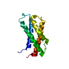
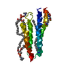
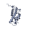
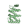
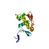
Signaling by BMP / Ribosome Quality Control (RQC) complex extracts and degrades nascent peptide / ZNF598 and the Ribosome-associated Quality Trigger (RQT) complex dissociate a ribosome stalled on a no-go mRNA / Oxygen-dependent proline hydroxylation of Hypoxia-inducible Factor Alpha / Regulation of TNFR1 signaling / Neddylation / Synthesis of active ubiquitin: roles of E1 and E2 enzymes / Downregulation of SMAD2/3:SMAD4 transcriptional activity / Ovarian tumor domain proteases / Antigen processing: Ubiquitination & Proteasome degradation ...Signaling by BMP / Ribosome Quality Control (RQC) complex extracts and degrades nascent peptide / ZNF598 and the Ribosome-associated Quality Trigger (RQT) complex dissociate a ribosome stalled on a no-go mRNA / Oxygen-dependent proline hydroxylation of Hypoxia-inducible Factor Alpha / Regulation of TNFR1 signaling / Neddylation / Synthesis of active ubiquitin: roles of E1 and E2 enzymes / Downregulation of SMAD2/3:SMAD4 transcriptional activity / Ovarian tumor domain proteases / Antigen processing: Ubiquitination & Proteasome degradation / programmed cell death involved in cell development / E2 ubiquitin-conjugating enzyme / ubiquitin conjugating enzyme activity / protein polyubiquitination / ubiquitin-protein transferase activity / chromosome / ubiquitin-dependent protein catabolic process / protein ubiquitination / ubiquitin protein ligase binding / ATP binding / nucleus / cytoplasm Similarity search - Function
Ubiquitin Conjugating Enzyme / Ubiquitin Conjugating Enzyme / Ubiquitin-conjugating enzyme, active site / Ubiquitin-conjugating (UBC) active site signature. / Ubiquitin-conjugating enzyme E2 / Ubiquitin-conjugating enzyme / Ubiquitin-conjugating (UBC) core domain profile. / Ubiquitin-conjugating enzyme E2, catalytic domain homologues / Ubiquitin-conjugating enzyme/RWD-like / Roll / Alpha Beta Similarity search - Domain/homology
In the structure databanks used in Yorodumi, some data are registered as the other names, "COVID-19 virus" and "2019-nCoV". Here are the details of the virus and the list of structure data.
Jan 31, 2019. EMDB accession codes are about to change! (news from PDBe EMDB page)
EMDB accession codes are about to change! (news from PDBe EMDB page)
The allocation of 4 digits for EMDB accession codes will soon come to an end. Whilst these codes will remain in use, new EMDB accession codes will include an additional digit and will expand incrementally as the available range of codes is exhausted. The current 4-digit format prefixed with “EMD-” (i.e. EMD-XXXX) will advance to a 5-digit format (i.e. EMD-XXXXX), and so on. It is currently estimated that the 4-digit codes will be depleted around Spring 2019, at which point the 5-digit format will come into force.
The EM Navigator/Yorodumi systems omit the EMD- prefix.
Related info.:Q: What is EMD? / ID/Accession-code notation in Yorodumi/EM Navigator
Yorodumi is a browser for structure data from EMDB, PDB, SASBDB, etc.
This page is also the successor to EM Navigator detail page, and also detail information page/front-end page for Omokage search.
The word "yorodu" (or yorozu) is an old Japanese word meaning "ten thousand". "mi" (miru) is to see.
Related info.:EMDB / PDB / SASBDB / Comparison of 3 databanks / Yorodumi Search / Aug 31, 2016. New EM Navigator & Yorodumi / Yorodumi Papers / Jmol/JSmol / Function and homology information / Changes in new EM Navigator and Yorodumi
 Movie
Movie Controller

 Yorodumi
 Open data
 Basic information
 Components
 Keywords
 Function and homology information
 X-RAY DIFFRACTION /
  Authors
 Citation
 Structure visualization
 Downloads & links
 Other downloads
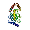


 PDBj
PDBj

 Assembly


 Mass: 35.453 Da / Num. of mol.: 6 / Source method: obtained synthetically / Formula: Cl
 Mass: 22.990 Da / Num. of mol.: 1 / Source method: obtained synthetically / Formula: Na
 Mass: 90.121 Da / Num. of mol.: 2 / Source method: obtained synthetically / Formula: C4H10O2
 Num. of mol.: 10 / Source method: obtained synthetically
 Sample preparation
 / Beamline: 22-ID / Wavelength: 0.97 Å
 Processing