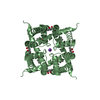
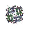
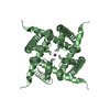
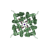
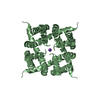
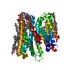
Journal: Sci Rep / Year: 2017 Title: Dead-end complex, lipid interactions and catalytic mechanism of microsomal glutathione transferase 1, an electron crystallography and mutagenesis investigation. Authors: Qie Kuang / Pasi Purhonen / Johan Ålander / Richard Svensson / Veronika Hoogland / Jens Winerdal / Linda Spahiu / Astrid Ottosson-Wadlund / Caroline Jegerschöld / Ralf Morgenstern / Hans Hebert / Abstract: Microsomal glutathione transferase 1 (MGST1) is a detoxification enzyme belonging to the Membrane Associated Proteins in Eicosanoid and Glutathione Metabolism (MAPEG) superfamily. Here we have used ...Microsomal glutathione transferase 1 (MGST1) is a detoxification enzyme belonging to the Membrane Associated Proteins in Eicosanoid and Glutathione Metabolism (MAPEG) superfamily. Here we have used electron crystallography of two-dimensional crystals in order to determine an atomic model of rat MGST1 in a lipid environment. The model comprises 123 of the 155 amino acid residues, two structured phospholipid molecules, two aliphatic chains and one glutathione (GSH) molecule. The functional unit is a homotrimer centered on the crystallographic three-fold axes of the unit cell. The GSH substrate binds in an extended conformation at the interface between two subunits of the trimer supported by new in vitro mutagenesis data. Mutation of Arginine 130 to alanine resulted in complete loss of activity consistent with a role for Arginine 130 in stabilizing the strongly nucleophilic GSH thiolate required for catalysis. Based on the new model and an electron diffraction data set from crystals soaked with trinitrobenzene, that forms a dead-end Meisenheimer complex with GSH, a difference map was calculated. The map reveals side chain movements opening a cavity that defines the second substrate site.
History
Deposition
Feb 20, 2016
Deposition site: RCSB / Processing site: PDBE
Revision 1.0
Jul 12, 2017
Provider: repository / Type: Initial release
Revision 1.0
Jul 12, 2017
Data content type: EM metadata / Data content type: EM metadata / Provider: repository / Type: Initial release
Revision 1.0
Jul 12, 2017
Data content type: Image / Data content type: Image / Provider: repository / Type: Initial release
Revision 1.0
Jul 12, 2017
Data content type: Primary map / Data content type: Primary map / Provider: repository / Type: Initial release
Revision 1.0
Jul 12, 2017
Data content type: EM metadata / Data content type: EM metadata / Provider: repository / Type: Initial release
Revision 1.0
Jul 12, 2017
Data content type: Image / Data content type: Image / Provider: repository / Type: Initial release
Revision 1.0
Jul 12, 2017
Data content type: Primary map / Data content type: Primary map / Provider: repository / Type: Initial release
Data content type: EM metadata / Data content type: EM metadata / EM metadata / Group: Database references / Experimental summary / Data content type: EM metadata / EM metadata / EM metadata / Category: database_2 / em_admin / em_author_list / Data content type: EM metadata / EM metadata / EM metadata Item: _database_2.pdbx_DOI / _database_2.pdbx_database_accession / _em_admin.last_update
-
Structure visualization
Movie
Biological unit as author_and_software_defined_assembly
Mass: 256.424 Da / Num. of mol.: 2 / Source method: obtained synthetically / Formula: C16H32O2
Has protein modification
N
-
Experimental details
-
Experiment
Experiment
Method: ELECTRON CRYSTALLOGRAPHY
EM experiment
Aggregation state: 2D ARRAY / 3D reconstruction method: electron crystallography
Crystal symmetry
∠γ: 120 ° / A: 81.8 Å / B: 81.8 Å / C: 100 Å / Space group name H-M: P6
-
Sample preparation
Component
Name: Complex between microsomal glutathione transferase 1 and glutathione Type: COMPLEX / Entity ID: #1 / Source: RECOMBINANT
Molecular weight
Value: 52940 MDa / Experimental value: NO
Source (natural)
Organism: Rattus norvegicus (Norway rat)
Source (recombinant)
Organism: Escherichia coli (E. coli) / Plasmid: pSP19T7LT
EM crystal formation
Details: Dialysis of protein solubulized in Triton X-100 / Lipid mixture: bovine liver lecithin / Lipid protein ratio: 3 / Temperature: 293 K / Time: 7 DAY
Buffer solution
pH: 7.4
Specimen
Embedding applied: YES / Shadowing applied: NO / Staining applied: NO / Vitrification applied: YES
EM embedding
Material: trehalose
Vitrification
Cryogen name: NITROGEN
-
Data collection
Microscopy
Model: JEOL 2100F
Electron gun
Electron source: FIELD EMISSION GUN / Accelerating voltage: 200 kV / Illumination mode: FLOOD BEAM
Electron lens
Mode: DIFFRACTION
Image recording
Electron dose: 1 e/Å2 / Film or detector model: TVIPS TEMCAM-F415 (4k x 4k) / Num. of diffraction images: 225
EM diffraction
Camera length: 200 mm
EM diffraction stats
Fourier space coverage: 82.5 % / High resolution: 3.5 Å / Num. of intensities measured: 97987 / Num. of structure factors: 6314 / Phase error: 1.0E-5 ° / Phase residual: 1.0E-5 ° / Phase error rejection criteria: 0 / Rmerge: 0.396 / Rsym: 0.126
 Movie
Movie Controller


 Open data
 Basic information
 Components
 Keywords
 Function and homology information
 Authors
 Sweden, 2items
  Citation
 Structure visualization
 Downloads & links
 Other downloads

 PDBj
PDBj


 Assembly
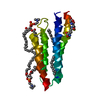


 Mass: 307.323 Da / Num. of mol.: 1 / Source method: obtained synthetically / Formula: C10H17N3O6S
 Mass: 790.145 Da / Num. of mol.: 2 / Source method: obtained synthetically / Formula: C44H88NO8P / Comment: phospholipid*YM
 Mass: 256.424 Da / Num. of mol.: 2 / Source method: obtained synthetically / Formula: C16H32O2
 Sample preparation
 FIELD EMISSION GUN / Accelerating voltage: 200 kV / Illumination mode: FLOOD BEAM
 Processing