 Movie
Movie Controller

[English] 日本語
 Yorodumi
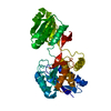
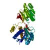
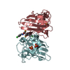
- PDB-1z18: Crystal structure analysis of periplasmic Leu/Ile/Val-binding pro... -
+ Open data

- Basic information
| Entry | Database: PDB / ID: 1z18 | ||||||
|---|---|---|---|---|---|---|---|
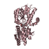
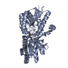
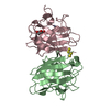
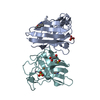
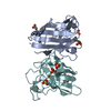
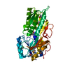
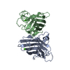
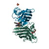
| Title | Crystal structure analysis of periplasmic Leu/Ile/Val-binding protein with bound valine | ||||||
 Components Components | Leu/Ile/Val-binding protein | ||||||
 Keywords Keywords | TRANSPORT PROTEIN / periplasmic binding proteins / alpha-beta fold / aliphatic amino acid binding protein | ||||||
| Function / homology |  Function and homology information Function and homology informationbranched-chain amino acid transport / valine transport / isoleucine transport / L-leucine transport / amino acid transport / ATP-binding cassette (ABC) transporter complex, substrate-binding subunit-containing / outer membrane-bounded periplasmic space / periplasmic space / membrane Similarity search - Function | ||||||
| Biological species |  | ||||||
| Method |  X-RAY DIFFRACTION / MOLECULAR REPLACEMENT / Resolution: 2.1 Å X-RAY DIFFRACTION / MOLECULAR REPLACEMENT / Resolution: 2.1 Å | ||||||
 Authors Authors | Trakhanov, S.D. / Vyas, N.K. / Kristensen, D.M. / Ma, J. / Quiocho, F.A. | ||||||
 Citation Citation | Journal: Biochemistry / Year: 2005 Title: Ligand-free and -bound structures of the binding protein (LivJ) of the Escherichia coli ABC leucine/isoleucine/valine transport system: trajectory and dynamics of the interdomain rotation and ligand specificity. Authors: Trakhanov, S.D. / Vyas, N.K. / Luecke, H. / Kristensen, D.M. / Ma, J. / Quiocho, F.A. #1: Journal: J.Mol.Biol. / Year: 1989Title: Periplasmic binding protein structure and function. Refined X-ray structures of the leucine/isoleucine/valine-binding protein and its complex with leucine. Authors: Sack, J.S. / Saper, M.A. / Quiocho, F.A. #2: Journal: J.Mol.Biol. / Year: 1989Title: Structure of the L-leucine-binding protein refined at 2.4 A resolution and comparison with the Leu/Ile/Val-binding protein structure. Authors: Sack, J.S. / Trakhanov, S.D. / Tsigannik, I.H. / Quiocho, F.A. | ||||||
| History |
|
- Structure visualization
| Structure viewer | Molecule: MolmilJmol/JSmol |
|---|
- Downloads & links
-Download
| PDBx/mmCIF format | 1z18.cif.gz | 81.3 KB | Display | PDBx/mmCIF format |
|---|---|---|---|---|
| PDB format | pdb1z18.ent.gz | 60.8 KB | Display | PDB format |
| PDBx/mmJSON format | 1z18.json.gz | Tree view | PDBx/mmJSON format | |
| Others |  Other downloads Other downloads |
-Validation report
| Arichive directory | https://data.pdbj.org/pub/pdb/validation_reports/z1/1z18ftp://data.pdbj.org/pub/pdb/validation_reports/z1/1z18 | HTTPS FTP |
|---|
-Related structure data
| Related structure data |  1z15C 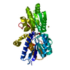 1z16SC 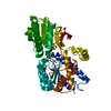 1z17C C: citing same article ( S: Starting model for refinement |
|---|---|
| Similar structure data |

-Links
 PDBj
PDBj
- Assembly
| Deposited unit | 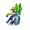
| ||||||||
|---|---|---|---|---|---|---|---|---|---|
| 1 |
| ||||||||
| Unit cell |
|
-Components
| #1: Protein | Mass: 36812.395 Da / Num. of mol.: 1 / Fragment: matured protein (residues 24-367) / Source method: isolated from a natural source / Source: (natural) | ||||||
|---|---|---|---|---|---|---|---|
| #2: Chemical |   Mass: 112.411 Da / Num. of mol.: 3 / Source method: obtained synthetically / Formula: Cd Mass: 112.411 Da / Num. of mol.: 3 / Source method: obtained synthetically / Formula: Cd#3: Chemical | ChemComp-VAL / |   Type: L-peptide linking / Mass: 117.146 Da / Num. of mol.: 1 / Source method: obtained synthetically / Formula: C5H11NO2 Type: L-peptide linking / Mass: 117.146 Da / Num. of mol.: 1 / Source method: obtained synthetically / Formula: C5H11NO2#4: Water | ChemComp-HOH / |  Mass: 18.015 Da / Num. of mol.: 195 / Source method: isolated from a natural source / Formula: H2O Mass: 18.015 Da / Num. of mol.: 195 / Source method: isolated from a natural source / Formula: H2OHas protein modification | Y | |
-Experimental details
-Experiment
| Experiment | Method: X-RAY DIFFRACTION / Number of used crystals: 1 |
|---|
- Sample preparation
| Crystal | Density Matthews: 2.28 Å3/Da / Density % sol: 45 % |
|---|---|
| Crystal grow | Temperature: 293 K / Method: vapor diffusion, hanging drop / pH: 5.5 Details: 25% (v/v) PEG-400, 0.1 M succinic acid, 50 mM CdSo4 and 50 mM Na-cacodylate, pH 5.5, VAPOR DIFFUSION, HANGING DROP, temperature 293K |
-Data collection
| Diffraction | Mean temperature: 100 K |
|---|---|
| Diffraction source | Source: ROTATING ANODE / Type: RIGAKU RU200 / Wavelength: 1.5418 Å |
| Detector | Type: MACSCIENCE / Detector: IMAGE PLATE / Date: Apr 5, 1998 / Details: Graphite monochromator |
| Radiation | Monochromator: Graphite / Protocol: SINGLE WAVELENGTH / Monochromatic (M) / Laue (L): M / Scattering type: x-ray |
| Radiation wavelength | Wavelength: 1.5418 Å / Relative weight: 1 |
| Reflection | Resolution: 2.1→50 Å / Num. all: 45340 / Num. obs: 18896 / % possible obs: 92 % / Observed criterion σ(F): 1 / Observed criterion σ(I): 1 / Redundancy: 3 % / Biso Wilson estimate: 0.5 Å2 / Rmerge(I) obs: 0.129 / Rsym value: 0.129 / Net I/σ(I): 5.4 |
| Reflection shell | Resolution: 2.1→2.2 Å / Redundancy: 2 % / Rmerge(I) obs: 0.242 / Mean I/σ(I) obs: 2.2 / Num. unique all: 18896 / Rsym value: 0.242 / % possible all: 85 |
- Processing
| Software |
| ||||||||||||||||||||||||||||||||||||||||||||||||||||||||||||
|---|---|---|---|---|---|---|---|---|---|---|---|---|---|---|---|---|---|---|---|---|---|---|---|---|---|---|---|---|---|---|---|---|---|---|---|---|---|---|---|---|---|---|---|---|---|---|---|---|---|---|---|---|---|---|---|---|---|---|---|---|---|
| Refinement | Method to determine structure: MOLECULAR REPLACEMENT Starting model: liv-leu complex without bound leu and solvent, pdb entry 1Z16 Resolution: 2.1→29.21 Å / Rfactor Rfree error: 0.009 / Data cutoff high absF: 91698.33 / Data cutoff low absF: 0 / Isotropic thermal model: RESTRAINED / Cross valid method: THROUGHOUT / σ(F): 0 / σ(I): 0 / Stereochemistry target values: Engh & Huber
| ||||||||||||||||||||||||||||||||||||||||||||||||||||||||||||
| Solvent computation | Solvent model: FLAT MODEL / Bsol: 54.2742 Å2 / ksol: 0.381888 e/Å3 | ||||||||||||||||||||||||||||||||||||||||||||||||||||||||||||
| Displacement parameters | Biso mean: 13.3 Å2
| ||||||||||||||||||||||||||||||||||||||||||||||||||||||||||||
| Refine analyze |
| ||||||||||||||||||||||||||||||||||||||||||||||||||||||||||||
| Refinement step | Cycle: LAST / Resolution: 2.1→29.21 Å
| ||||||||||||||||||||||||||||||||||||||||||||||||||||||||||||
| Refine LS restraints |
| ||||||||||||||||||||||||||||||||||||||||||||||||||||||||||||
| LS refinement shell | Resolution: 2.1→2.23 Å / Rfactor Rfree error: 0.025 / Total num. of bins used: 6
| ||||||||||||||||||||||||||||||||||||||||||||||||||||||||||||
| Xplor file |
|
