 Movie
Movie Controller

[English] 日本語
 Yorodumi
- PDB-1yy8: Crystal structure of the Fab fragment from the monoclonal antibod... -
+ Open data

- Basic information
| Entry | Database: PDB / ID: 1yy8 | ||||||
|---|---|---|---|---|---|---|---|
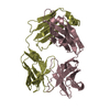
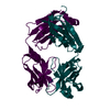
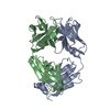
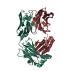
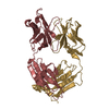
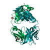
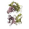
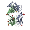
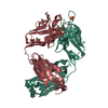
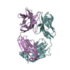
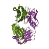
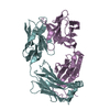
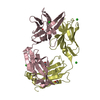
| Title | Crystal structure of the Fab fragment from the monoclonal antibody cetuximab/Erbitux/IMC-C225 | ||||||
 Components Components |
| ||||||
 Keywords Keywords | IMMUNE SYSTEM / FAB FRAGMENT / ANTIBODY DRUG / CANCER | ||||||
| Function / homology | Immunoglobulins / Immunoglobulin-like / Sandwich / Mainly Beta Function and homology information Function and homology information | ||||||
| Biological species |   Homo sapiens (human) Homo sapiens (human) | ||||||
| Method |  X-RAY DIFFRACTION / SYNCHROTRON / MOLECULAR REPLACEMENT / Resolution: 2 Å X-RAY DIFFRACTION / SYNCHROTRON / MOLECULAR REPLACEMENT / Resolution: 2 Å | ||||||
 Authors Authors | Li, S. / Schmitz, K.R. / Jeffrey, P.D. / Wiltzius, J.J.W. / Kussie, P. / Ferguson, K.M. | ||||||
 Citation Citation | Journal: Cancer Cell / Year: 2005 Title: Structural basis for inhibition of the epidermal growth factor receptor by cetuximab Authors: Li, S. / Schmitz, K.R. / Jeffrey, P.D. / Wiltzius, J.J.W. / Kussie, P. / Ferguson, K.M. | ||||||
| History |
|
- Structure visualization
| Structure viewer | Molecule: MolmilJmol/JSmol |
|---|
- Downloads & links
-Download
| PDBx/mmCIF format | 1yy8.cif.gz | 179.2 KB | Display | PDBx/mmCIF format |
|---|---|---|---|---|
| PDB format | pdb1yy8.ent.gz | 143.4 KB | Display | PDB format |
| PDBx/mmJSON format | 1yy8.json.gz | Tree view | PDBx/mmJSON format | |
| Others |  Other downloads Other downloads |
-Validation report
| Arichive directory | https://data.pdbj.org/pub/pdb/validation_reports/yy/1yy8ftp://data.pdbj.org/pub/pdb/validation_reports/yy/1yy8 | HTTPS FTP |
|---|
-Related structure data
| Related structure data |  1yy9C 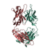 1ad9S 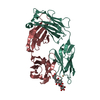 1ibgS C: citing same article ( S: Starting model for refinement |
|---|---|
| Similar structure data |


-Links
 PDBj
PDBj


- Assembly
| Deposited unit | 
| ||||||||||
|---|---|---|---|---|---|---|---|---|---|---|---|
| 1 | 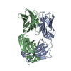
| ||||||||||
| 2 | 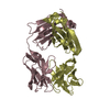
| ||||||||||
| Unit cell |
|
-Components
| #1: Antibody | Mass: 23287.705 Da / Num. of mol.: 2 Source method: isolated from a genetically manipulated source Source: (gene. exp.) Mus musculus, Homo sapiens / Genus: Mus, Homo / Species: , / Strain: , / Plasmid: pdHL2 / Cell (production host): mouse myeloma cell line / Production host: #2: Antibody | Mass: 23725.504 Da / Num. of mol.: 2 Source method: isolated from a genetically manipulated source Source: (gene. exp.) Mus musculus, Homo sapiens / Genus: Mus, Homo / Species: , / Strain: , / Plasmid: pdHL2 / Cell (production host): mouse myeloma cell line / Production host: #3: Water | ChemComp-HOH / |  Mass: 18.015 Da / Num. of mol.: 354 / Source method: isolated from a natural source / Formula: H2O Mass: 18.015 Da / Num. of mol.: 354 / Source method: isolated from a natural source / Formula: H2OHas protein modification | Y | |
|---|
-Experimental details
-Experiment
| Experiment | Method: X-RAY DIFFRACTION / Number of used crystals: 1 |
|---|
- Sample preparation
| Crystal | Density Matthews: 2.87 Å3/Da / Density % sol: 56.7 % |
|---|---|
| Crystal grow | Method: vapor diffusion, hanging drop / pH: 6.5 Details: Ammonium sulfate, pH 6.5, VAPOR DIFFUSION, HANGING DROP |
-Data collection
| Diffraction | Mean temperature: 100 K |
|---|---|
| Diffraction source | Source: SYNCHROTRON / Site: NSLS  / Beamline: X9B / Beamline: X9B |
| Detector | Type: MARRESEARCH / Detector: CCD |
| Radiation | Protocol: SINGLE WAVELENGTH / Monochromatic (M) / Laue (L): M / Scattering type: x-ray |
| Radiation wavelength | Relative weight: 1 |
| Reflection | Resolution: 2→50 Å / Num. all: 73414 / Num. obs: 73414 / % possible obs: 95.6 % / Observed criterion σ(I): 0.5 / Redundancy: 8.6 % / Rmerge(I) obs: 0.073 / Rsym value: 0.073 / Net I/σ(I): 12.7 |
| Reflection shell | Resolution: 2→2.07 Å / Rmerge(I) obs: 0.297 / Mean I/σ(I) obs: 5.5 / % possible all: 0.968 |
- Processing
| Software |
| ||||||||||||||||||||||||||||
|---|---|---|---|---|---|---|---|---|---|---|---|---|---|---|---|---|---|---|---|---|---|---|---|---|---|---|---|---|---|
| Refinement | Method to determine structure: MOLECULAR REPLACEMENT Starting model: pdb ids 1IBG, 1AD9 Resolution: 2→15 Å / Data cutoff high absF: 10000 / Data cutoff low absF: 0 / Cross valid method: THROUGHOUT / σ(F): 0
| ||||||||||||||||||||||||||||
| Displacement parameters |
| ||||||||||||||||||||||||||||
| Refinement step | Cycle: LAST / Resolution: 2→15 Å
| ||||||||||||||||||||||||||||
| Refine LS restraints |
|
