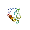
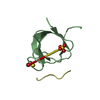
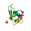
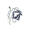
[English] 日本語
Yorodumi
- PDB-1ywo: Phospholipase Cgamma1 SH3 in complex with a SLP-76 motif -

+
Open data


ID or keywords:

Loading...

-
Basic information

Entry
Database: PDB / ID: 1ywo
TitlePhospholipase Cgamma1 SH3 in complex with a SLP-76 motif
Components
  • 1-phosphatidylinositol-4,5-bisphosphate phosphodiesterase gamma 1
  • Lymphocyte cytosolic protein 2
KeywordsHYDROLASE / SIGNALING PROTEIN / SH3 / Phospholipase C-gamma1 / SLP-76 / SH2 domain-containing leukocyte phosphoprotein of 76 kD
Function / homology
Function and homology information


TCR signalosome / PECAM1 interactions / EGFR interacts with phospholipase C-gamma / Activated NTRK2 signals through PLCG1 / Activated NTRK3 signals through PLCG1 / phosphatidylinositol catabolic process / Phospholipase C-mediated cascade: FGFR1 / Phospholipase C-mediated cascade; FGFR3 / Phospholipase C-mediated cascade; FGFR4 / FCERI mediated Ca+2 mobilization ...TCR signalosome / PECAM1 interactions / EGFR interacts with phospholipase C-gamma / Activated NTRK2 signals through PLCG1 / Activated NTRK3 signals through PLCG1 / phosphatidylinositol catabolic process / Phospholipase C-mediated cascade: FGFR1 / Phospholipase C-mediated cascade; FGFR3 / Phospholipase C-mediated cascade; FGFR4 / FCERI mediated Ca+2 mobilization / ISG15 antiviral mechanism / Generation of second messenger molecules / Phospholipase C-mediated cascade; FGFR2 / Downstream signal transduction / Signaling by ALK / Role of phospholipids in phagocytosis / inositol trisphosphate biosynthetic process / DAP12 signaling / VEGFR2 mediated cell proliferation / calcium-dependent phospholipase C activity / Synthesis of IP3 and IP4 in the cytosol / RET signaling / inositol trisphosphate metabolic process / response to curcumin / mast cell activation / phosphoinositide phospholipase C / FCERI mediated MAPK activation / response to gravity / phosphatidylinositol metabolic process / phosphatidylinositol phospholipase C activity / COP9 signalosome / phospholipase C activity / neurotrophin TRKA receptor binding / positive regulation of endothelial cell apoptotic process / phosphatidylinositol-mediated signaling / positive regulation of vascular endothelial cell proliferation / clathrin-coated vesicle / positive regulation of epithelial cell migration / plasma membrane raft / Generation of second messenger molecules / glutamate receptor binding / positive regulation of blood vessel endothelial cell migration / positive regulation of protein kinase activity / release of sequestered calcium ion into cytosol / cellular response to epidermal growth factor stimulus / ruffle / GPVI-mediated activation cascade / response to organonitrogen compound / FCERI mediated Ca+2 mobilization / guanyl-nucleotide exchange factor activity / positive regulation of release of sequestered calcium ion into cytosol / cell projection / calcium-mediated signaling / FCERI mediated MAPK activation / phosphoprotein binding / modulation of chemical synaptic transmission / Schaffer collateral - CA1 synapse / response to hydrogen peroxide / insulin receptor binding / epidermal growth factor receptor signaling pathway / receptor tyrosine kinase binding / ruffle membrane / cell surface receptor protein tyrosine kinase signaling pathway / positive regulation of angiogenesis / calcium ion transport / cell-cell junction / cell migration / DAP12 signaling / lamellipodium / T cell receptor signaling pathway / in utero embryonic development / intracellular signal transduction / immune response / glutamatergic synapse / calcium ion binding / protein kinase binding / plasma membrane / cytosol / cytoplasm
Similarity search - Function
1-phosphatidylinositol 4,5-bisphosphate phosphodiesterase gamma-1, SH3 domain / Phosphatidylinositol-4, 5-bisphosphate phosphodiesterase gamma / PLC-gamma, C-terminal SH2 domain / PLC-gamma, N-terminal SH2 domain / Phosphoinositide phospholipase C family / Phospholipase C, phosphatidylinositol-specific, Y domain / Phosphatidylinositol-specific phospholipase C, Y domain / Phosphatidylinositol-specific phospholipase Y-box domain profile. / Phospholipase C, catalytic domain (part); domain Y / Phosphatidylinositol-specific phospholipase X-box domain profile. ...1-phosphatidylinositol 4,5-bisphosphate phosphodiesterase gamma-1, SH3 domain / Phosphatidylinositol-4, 5-bisphosphate phosphodiesterase gamma / PLC-gamma, C-terminal SH2 domain / PLC-gamma, N-terminal SH2 domain / Phosphoinositide phospholipase C family / Phospholipase C, phosphatidylinositol-specific, Y domain / Phosphatidylinositol-specific phospholipase C, Y domain / Phosphatidylinositol-specific phospholipase Y-box domain profile. / Phospholipase C, catalytic domain (part); domain Y / Phosphatidylinositol-specific phospholipase X-box domain profile. / Phosphatidylinositol-specific phospholipase C, X domain / Phosphatidylinositol-specific phospholipase C, X domain / Phospholipase C, catalytic domain (part); domain X / PLC-like phosphodiesterase, TIM beta/alpha-barrel domain superfamily / C2 domain / Protein kinase C conserved region 2 (CalB) / C2 domain / C2 domain profile. / SAM domain (Sterile alpha motif) / SH3 Domains / Sterile alpha motif. / Sterile alpha motif domain / PH domain / Sterile alpha motif/pointed domain superfamily / C2 domain superfamily / PH domain profile. / Pleckstrin homology domain. / Pleckstrin homology domain / SH3 domain / SH2 domain / Src homology 2 (SH2) domain profile. / Src homology 2 domains / SH2 domain / Src homology 3 domains / SH3 type barrels. / EF-Hand 1, calcium-binding site / EF-hand calcium-binding domain. / SH2 domain superfamily / EF-hand calcium-binding domain profile. / SH3-like domain superfamily / Src homology 3 (SH3) domain profile. / SH3 domain / EF-hand domain / EF-hand domain pair / PH-like domain superfamily / Roll / Mainly Beta
Similarity search - Domain/homology
1-phosphatidylinositol 4,5-bisphosphate phosphodiesterase gamma-1 / Lymphocyte cytosolic protein 2
Similarity search - Component
Biological speciesRattus norvegicus (Norway rat)
Homo sapiens (human)
MethodX-RAY DIFFRACTION / SYNCHROTRON / MOLECULAR REPLACEMENT / Resolution: 1.81 Å
AuthorsDeng, L. / Velikovsky, C.A. / Swaminathan, C.P. / Cho, S. / Mariuzza, R.A.
CitationJournal: J.Mol.Biol. / Year: 2005
Title: Structural Basis for Recognition of the T Cell Adaptor Protein SLP-76 by the SH3 Domain of Phospholipase Cgamma1
Authors: Deng, L. / Velikovsky, C.A. / Swaminathan, C.P. / Cho, S. / Mariuzza, R.A.
History
DepositionFeb 18, 2005Deposition site: RCSB / Processing site: RCSB
Revision 1.0Aug 16, 2005Provider: repository / Type: Initial release
Revision 1.1Apr 30, 2008Group: Version format compliance
Revision 1.2Jul 13, 2011Group: Advisory / Version format compliance
Revision 1.3Oct 20, 2021Group: Database references / Category: database_2 / struct_ref_seq_dif
Item: _database_2.pdbx_DOI / _database_2.pdbx_database_accession / _struct_ref_seq_dif.details
Revision 1.4Feb 14, 2024Group: Data collection / Category: chem_comp_atom / chem_comp_bond

-
Structure visualization

Structure viewerMolecule:
MolmilJmol/JSmol

Downloads & links

-
Assembly

Deposited unit
A: 1-phosphatidylinositol-4,5-bisphosphate phosphodiesterase gamma 1
P: Lymphocyte cytosolic protein 2


Theoretical massNumber of molelcules
Total (without water)8,5622
Polymers8,5622
Non-polymers00
Water1,20767
1


  • Idetical with deposited unit
  • defined by author
TypeNameSymmetry operationNumber
identity operation1_555x,y,z1
Unit cell
Length a, b, c (Å)46.723, 46.723, 58.112
Angle α, β, γ (deg.)90.00, 90.00, 90.00
Int Tables number96
Space group name H-MP43212

-
Components

#1: Protein 1-phosphatidylinositol-4,5-bisphosphate phosphodiesterase gamma 1 / Phosphoinositide phospholipase C / PLC-gamma-1 / Phospholipase C-gamma-1 / PLC-II / PLC-148


Mass: 7414.259 Da / Num. of mol.: 1 / Mutation: C5S
Source method: isolated from a genetically manipulated source
Source: (gene. exp.) Rattus norvegicus (Norway rat) / Gene: Plcg1 / Plasmid: pGEX-4T-1 / Species (production host): Escherichia coli / Production host: Escherichia coli BL21(DE3) (bacteria) / Strain (production host): BL21(DE3)
References: UniProt: P10686, phosphoinositide phospholipase C
#2: Protein/peptide Lymphocyte cytosolic protein 2 / / SH2 domain-containing leucocyte protein of 76 kDa / SLP-76 tyrosine phosphoprotein / SLP76


Mass: 1147.367 Da / Num. of mol.: 1 / Source method: isolated from a natural source / Source: (natural) Homo sapiens (human) / References: UniProt: Q13094
#3: Water ChemComp-HOH / water / Water


Mass: 18.015 Da / Num. of mol.: 67 / Source method: isolated from a natural source / Formula: H2O

-
Experimental details

-
Experiment

ExperimentMethod: X-RAY DIFFRACTION / Number of used crystals: 1

-
Sample preparation

CrystalDensity Matthews: 1.9 Å3/Da / Density % sol: 33.2 %
Crystal growTemperature: 298 K / Method: vapor diffusion, hanging drop / pH: 7
Details: malonate , pH 7.0, VAPOR DIFFUSION, HANGING DROP, temperature 298.0K

-
Data collection

DiffractionMean temperature: 100 K
Diffraction sourceSource: SYNCHROTRON / Site: NSLS / Beamline: X29A / Wavelength: 1.009 Å
DetectorType: ADSC QUANTUM 315 / Detector: CCD / Date: Nov 9, 2004
RadiationProtocol: SINGLE WAVELENGTH / Monochromatic (M) / Laue (L): M / Scattering type: x-ray
Radiation wavelengthWavelength: 1.009 Å / Relative weight: 1
ReflectionResolution: 1.81→50 Å / Num. obs: 6149 / Rmerge(I) obs: 0.097 / Net I/σ(I): 21.7
Reflection shellResolution: 1.81→1.87 Å / Rmerge(I) obs: 0.35 / Mean I/σ(I) obs: 1.8 / % possible all: 85.8

-
Processing

Software
NameVersionClassification
REFMAC5.2.0003refinement
HKL-2000data reduction
SCALEPACKdata scaling
PHASESphasing
RefinementMethod to determine structure: MOLECULAR REPLACEMENT / Resolution: 1.81→36.42 Å / Cor.coef. Fo:Fc: 0.963 / Cor.coef. Fo:Fc free: 0.931 / SU B: 5.412 / SU ML: 0.095 / TLS residual ADP flag: LIKELY RESIDUAL / Cross valid method: THROUGHOUT / ESU R: 0.135 / ESU R Free: 0.133 / Stereochemistry target values: MAXIMUM LIKELIHOOD
RfactorNum. reflection% reflectionSelection details
Rfree0.22129 279 4.6 %RANDOM
Rwork0.17063 ---
obs0.17285 5831 96.74 %-
Solvent computationIon probe radii: 0.8 Å / Shrinkage radii: 0.8 Å / VDW probe radii: 1.2 Å / Solvent model: MASK
Displacement parametersBiso mean: 23.064 Å2
Baniso -1Baniso -2Baniso -3
1-0.54 Å20 Å20 Å2
2--0.54 Å20 Å2
3----1.08 Å2
Refinement stepCycle: LAST / Resolution: 1.81→36.42 Å
ProteinNucleic acidLigandSolventTotal
Num. atoms536 0 0 67 603
Refine LS restraints
Refine-IDTypeDev idealDev ideal targetNumber
X-RAY DIFFRACTIONr_bond_refined_d0.0140.022563
X-RAY DIFFRACTIONr_bond_other_d
X-RAY DIFFRACTIONr_angle_refined_deg1.4051.946761
X-RAY DIFFRACTIONr_angle_other_deg
X-RAY DIFFRACTIONr_dihedral_angle_1_deg6.285563
X-RAY DIFFRACTIONr_dihedral_angle_2_deg43.84424.51631
X-RAY DIFFRACTIONr_dihedral_angle_3_deg14.641597
X-RAY DIFFRACTIONr_dihedral_angle_4_deg18.613154
X-RAY DIFFRACTIONr_chiral_restr0.0950.272
X-RAY DIFFRACTIONr_gen_planes_refined0.0070.02447
X-RAY DIFFRACTIONr_gen_planes_other
X-RAY DIFFRACTIONr_nbd_refined0.2130.2226
X-RAY DIFFRACTIONr_nbd_other
X-RAY DIFFRACTIONr_nbtor_refined0.3070.2375
X-RAY DIFFRACTIONr_nbtor_other
X-RAY DIFFRACTIONr_xyhbond_nbd_refined0.1890.245
X-RAY DIFFRACTIONr_xyhbond_nbd_other
X-RAY DIFFRACTIONr_metal_ion_refined
X-RAY DIFFRACTIONr_metal_ion_other
X-RAY DIFFRACTIONr_symmetry_vdw_refined0.2240.243
X-RAY DIFFRACTIONr_symmetry_vdw_other
X-RAY DIFFRACTIONr_symmetry_hbond_refined0.2560.211
X-RAY DIFFRACTIONr_symmetry_hbond_other
X-RAY DIFFRACTIONr_symmetry_metal_ion_refined
X-RAY DIFFRACTIONr_symmetry_metal_ion_other
X-RAY DIFFRACTIONr_mcbond_it1.6672331
X-RAY DIFFRACTIONr_mcbond_other
X-RAY DIFFRACTIONr_mcangle_it2.5443527
X-RAY DIFFRACTIONr_scbond_it1.7392275
X-RAY DIFFRACTIONr_scangle_it2.6453234
X-RAY DIFFRACTIONr_rigid_bond_restr
X-RAY DIFFRACTIONr_sphericity_free
X-RAY DIFFRACTIONr_sphericity_bonded
LS refinement shellResolution: 1.805→1.852 Å / Total num. of bins used: 20
RfactorNum. reflection% reflection
Rfree0.373 15 -
Rwork0.269 327 -
obs--77.2 %
Refinement TLS params.Method: refined / Origin x: 20.4361 Å / Origin y: 11.5163 Å / Origin z: 17.5977 Å
111213212223313233
T-0.0418 Å20.0036 Å2-0.0041 Å2--0.0038 Å2-0.0233 Å2---0.023 Å2
L2.5795 °21.7207 °2-0.2661 °2-2.211 °2-0.7526 °2--4.0509 °2
S-0.019 Å °-0.0011 Å °0.1814 Å °-0.0076 Å °0.0482 Å °0.1718 Å °-0.0291 Å °-0.1294 Å °-0.0292 Å °

+
About Yorodumi

-
News

-
Feb 9, 2022. New format data for meta-information of EMDB entries

New format data for meta-information of EMDB entries

  • Version 3 of the EMDB header file is now the official format.
  • The previous official version 1.9 will be removed from the archive.

Related info.:EMDB header

External links:wwPDB to switch to version 3 of the EMDB data model

-
Aug 12, 2020. Covid-19 info

Covid-19 info

URL: https://pdbj.org/emnavi/covid19.php

New page: Covid-19 featured information page in EM Navigator.

Related info.:Covid-19 info / Mar 5, 2020. Novel coronavirus structure data

+
Mar 5, 2020. Novel coronavirus structure data

Novel coronavirus structure data

Related info.:Yorodumi Speices / Aug 12, 2020. Covid-19 info

External links:COVID-19 featured content - PDBj / Molecule of the Month (242):Coronavirus Proteases

+
Jan 31, 2019. EMDB accession codes are about to change! (news from PDBe EMDB page)

EMDB accession codes are about to change! (news from PDBe EMDB page)

  • The allocation of 4 digits for EMDB accession codes will soon come to an end. Whilst these codes will remain in use, new EMDB accession codes will include an additional digit and will expand incrementally as the available range of codes is exhausted. The current 4-digit format prefixed with “EMD-” (i.e. EMD-XXXX) will advance to a 5-digit format (i.e. EMD-XXXXX), and so on. It is currently estimated that the 4-digit codes will be depleted around Spring 2019, at which point the 5-digit format will come into force.
  • The EM Navigator/Yorodumi systems omit the EMD- prefix.

Related info.:Q: What is EMD? / ID/Accession-code notation in Yorodumi/EM Navigator

External links:EMDB Accession Codes are Changing Soon! / Contact to PDBj

+
Jul 12, 2017. Major update of PDB

Major update of PDB

  • wwPDB released updated PDB data conforming to the new PDBx/mmCIF dictionary.
  • This is a major update changing the version number from 4 to 5, and with Remediation, in which all the entries are updated.
  • In this update, many items about electron microscopy experimental information are reorganized (e.g. em_software).
  • Now, EM Navigator and Yorodumi are based on the updated data.

External links:wwPDB Remediation / Enriched Model Files Conforming to OneDep Data Standards Now Available in the PDB FTP Archive

-
Yorodumi

Thousand views of thousand structures

  • Yorodumi is a browser for structure data from EMDB, PDB, SASBDB, etc.
  • This page is also the successor to EM Navigator detail page, and also detail information page/front-end page for Omokage search.
  • The word "yorodu" (or yorozu) is an old Japanese word meaning "ten thousand". "mi" (miru) is to see.

Related info.:EMDB / PDB / SASBDB / Comparison of 3 databanks / Yorodumi Search / Aug 31, 2016. New EM Navigator & Yorodumi / Yorodumi Papers / Jmol/JSmol / Function and homology information / Changes in new EM Navigator and Yorodumi

Read more