 Movie
Movie Controller

[English] 日本語
 Yorodumi
- PDB-1ynt: Structure of the monomeric form of T. gondii SAG1 surface antigen... -
+ Open data

- Basic information
| Entry | Database: PDB / ID: 1ynt | ||||||
|---|---|---|---|---|---|---|---|
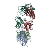
| Title | Structure of the monomeric form of T. gondii SAG1 surface antigen bound to a human Fab | ||||||
 Components Components |
| ||||||
 Keywords Keywords | IMMUNE SYSTEM / Toxoplasma gondii / recombinant SAG1 / conformational epitope | ||||||
| Function / homology |  Function and homology information Function and homology informationsymbiont-containing vacuole / IgG binding / immunoglobulin binding / membrane Similarity search - Function | ||||||
| Biological species |  Finegoldia magna (bacteria) Finegoldia magna (bacteria)  | ||||||
| Method |  X-RAY DIFFRACTION / SYNCHROTRON / MOLECULAR REPLACEMENT / Resolution: 3.1 Å X-RAY DIFFRACTION / SYNCHROTRON / MOLECULAR REPLACEMENT / Resolution: 3.1 Å | ||||||
 Authors Authors | Graille, M. / Stura, E.A. / Bossus, M. / Muller, B.H. / Letourneur, O. / Battail-Poirot, N. / Sibai, G. / Rolland, D. / Le Du, M.H. / Ducancel, F. | ||||||
 Citation Citation | Journal: J.Mol.Biol. / Year: 2005 Title: Crystal structure of the complex between the monomeric form of Toxoplasma gondii surface antigen 1 (SAG1) and a monoclonal antibody that mimics the human immune response Authors: Graille, M. / Stura, E.A. / Bossus, M. / Muller, B.H. / Letourneur, O. / Battail-Poirot, N. / Sibai, G. / Gauthier, M. / Rolland, D. / Le Du, M.H. / Ducancel, F. | ||||||
| History |
|
- Structure visualization
| Structure viewer | Molecule: MolmilJmol/JSmol |
|---|
- Downloads & links
-Download
| PDBx/mmCIF format | 1ynt.cif.gz | 282.3 KB | Display | PDBx/mmCIF format |
|---|---|---|---|---|
| PDB format | pdb1ynt.ent.gz | 222.6 KB | Display | PDB format |
| PDBx/mmJSON format | 1ynt.json.gz | Tree view | PDBx/mmJSON format | |
| Others |  Other downloads Other downloads |
-Validation report
| Arichive directory | https://data.pdbj.org/pub/pdb/validation_reports/yn/1yntftp://data.pdbj.org/pub/pdb/validation_reports/yn/1ynt | HTTPS FTP |
|---|
-Related structure data


-Links
 PDBj
PDBj

- Assembly
| Deposited unit | 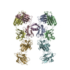
| ||||||||
|---|---|---|---|---|---|---|---|---|---|
| 1 |
| ||||||||
| Unit cell |
| ||||||||
| Details | The biological assembly is a Fab - antigen complex. Two are present in the asymetric unit |
-Components
| #1: Antibody | Mass: 23577.820 Da / Num. of mol.: 2 / Fragment: Fab fragment / Source method: isolated from a natural source / Source: (natural) #2: Antibody | Mass: 23568.344 Da / Num. of mol.: 2 / Fragment: Fab fragment / Source method: isolated from a natural source / Source: (natural) #3: Protein | | Mass: 6750.485 Da / Num. of mol.: 1 / Fragment: domain C* Source method: isolated from a genetically manipulated source Source: (gene. exp.) Finegoldia magna (bacteria) / Strain: ATCC 29328 / Plasmid: pKK223-3 / Production host: #4: Protein | Mass: 26640.217 Da / Num. of mol.: 2 / Fragment: residues in database 50-303 Source method: isolated from a genetically manipulated source Source: (gene. exp.)  Pichia pastoris (fungus) / References: UniProt: P13664 Pichia pastoris (fungus) / References: UniProt: P13664#5: Chemical |   Mass: 112.411 Da / Num. of mol.: 2 / Source method: obtained synthetically / Formula: Cd Mass: 112.411 Da / Num. of mol.: 2 / Source method: obtained synthetically / Formula: CdHas protein modification | Y | |
|---|
-Experimental details
-Experiment
| Experiment | Method: X-RAY DIFFRACTION / Number of used crystals: 1 |
|---|
- Sample preparation
| Crystal | Density Matthews: 2.8 Å3/Da / Density % sol: 57.985123 % |
|---|---|
| Crystal grow | Temperature: 291 K / Method: vapor diffusion, sitting drop / pH: 11 Details: 15% polyethylene glycol 3350, 0.25M NaCl, 150mM CAPS, pH 11, VAPOR DIFFUSION, SITTING DROP, temperature 291K |
-Data collection
| Diffraction | Mean temperature: 100 K |
|---|---|
| Diffraction source | Source: SYNCHROTRON / Site: ESRF  / Beamline: ID14-1 / Wavelength: 0.934 Å / Beamline: ID14-1 / Wavelength: 0.934 Å |
| Detector | Type: ADSC QUANTUM 4 / Detector: CCD / Date: Oct 29, 2001 |
| Radiation | Monochromator: Si 111 CHANNEL / Protocol: SINGLE WAVELENGTH / Monochromatic (M) / Laue (L): M / Scattering type: x-ray |
| Radiation wavelength | Wavelength: 0.934 Å / Relative weight: 1 |
| Reflection | Resolution: 3.1→40 Å / Num. all: 32176 / Num. obs: 31886 / % possible obs: 99.1 % / Observed criterion σ(F): 0 / Observed criterion σ(I): 0 / Rmerge(I) obs: 0.091 / Χ2: 1.241 |
| Reflection shell | Resolution: 3.1→3.19 Å / Rmerge(I) obs: 0.497 / Num. unique all: 2608 / Χ2: 0.866 / % possible all: 97.9 |
- Processing
| Software |
| ||||||||||||||||||||||||
|---|---|---|---|---|---|---|---|---|---|---|---|---|---|---|---|---|---|---|---|---|---|---|---|---|---|
| Refinement | Method to determine structure: MOLECULAR REPLACEMENT Starting model: PDB codes: 1F11, 1KB5, 1KZQ Resolution: 3.1→20 Å / Data cutoff high absF: 10000 / Data cutoff low absF: 0 / σ(F): 0 / Stereochemistry target values: Engh & Huber
| ||||||||||||||||||||||||
| Displacement parameters | Biso mean: 10 Å2
| ||||||||||||||||||||||||
| Refinement step | Cycle: LAST / Resolution: 3.1→20 Å
| ||||||||||||||||||||||||
| Refine LS restraints |
|
