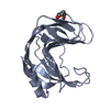
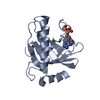
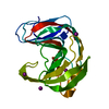
 Movie
Movie Controller


+ Open data

- Basic information
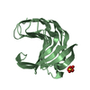
| Entry | Database: PDB / ID: 1yna | |||||||||
|---|---|---|---|---|---|---|---|---|---|---|
| Title | ENDO-1,4-BETA-XYLANASE, ROOM TEMPERATURE, PH 4.0 | |||||||||
 Components Components | ENDO-1,4-BETA-XYLANASE | |||||||||
 Keywords Keywords | HYDROLASE / XYLANASE | |||||||||
| Function / homology |  Function and homology information Function and homology informationendo-1,4-beta-xylanase / endo-1,4-beta-xylanase activity / xylan catabolic process Similarity search - Function | |||||||||
| Biological species |   Thermomyces lanuginosus (fungus) Thermomyces lanuginosus (fungus) | |||||||||
| Method |  X-RAY DIFFRACTION / MOLECULAR REPLACEMENT / Resolution: 1.55 Å X-RAY DIFFRACTION / MOLECULAR REPLACEMENT / Resolution: 1.55 Å | |||||||||
 Authors Authors | Gruber, K. / Kratky, C. | |||||||||
 Citation Citation | Journal: Biochemistry / Year: 1998 Title: Thermophilic xylanase from Thermomyces lanuginosus: high-resolution X-ray structure and modeling studies. Authors: Gruber, K. / Klintschar, G. / Hayn, M. / Schlacher, A. / Steiner, W. / Kratky, C. #1: Journal: J.Biotechnol. / Year: 1996Title: Three-Dimensional Structure of Endo-1,4-Beta-Xylanase II from Trichoderma Reesei: Two Conformational States in the Active Site Authors: Schlacher, A. / Holzmann, K. / Hayn, M. / Steiner, W. / Schwab, H. #2: Journal: Embo J. / Year: 1994Title: Three-Dimensional Structure of Endo-1,4-Beta-Xylanase II from Trichoderma Reesei: Two Conformational States in the Active Site Authors: Torronen, A. / Harkki, A. / Rouvinen, J. | |||||||||
| History |
|
- Structure visualization
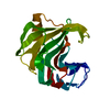
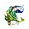
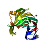
| Structure viewer | Molecule: MolmilJmol/JSmol |
|---|
- Downloads & links
-Download
| PDBx/mmCIF format | 1yna.cif.gz | 52.5 KB | Display | PDBx/mmCIF format |
|---|---|---|---|---|
| PDB format | pdb1yna.ent.gz | 37 KB | Display | PDB format |
| PDBx/mmJSON format | 1yna.json.gz | Tree view | PDBx/mmJSON format | |
| Others |  Other downloads Other downloads |
-Validation report
| Arichive directory | https://data.pdbj.org/pub/pdb/validation_reports/yn/1ynaftp://data.pdbj.org/pub/pdb/validation_reports/yn/1yna | HTTPS FTP |
|---|
-Related structure data
| Similar structure data |
|---|


-Links
 PDBj
PDBj
- Assembly
| Deposited unit | 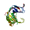
| ||||||||
|---|---|---|---|---|---|---|---|---|---|
| 1 |
| ||||||||
| Unit cell |
|
-Components
| #1: Protein | Mass: 21311.848 Da / Num. of mol.: 1 / Source method: isolated from a natural source / Source: (natural) Thermomyces lanuginosus (fungus) / Strain: TSIKLINSKY / References: UniProt: O43097, endo-1,4-beta-xylanase |
|---|---|
| #2: Water | ChemComp-HOH /  Mass: 18.015 Da / Num. of mol.: 138 / Source method: isolated from a natural source / Formula: H2O Mass: 18.015 Da / Num. of mol.: 138 / Source method: isolated from a natural source / Formula: H2O |
| Has protein modification | Y |
-Experimental details
-Experiment
| Experiment | Method: X-RAY DIFFRACTION / Number of used crystals: 1 |
|---|
- Sample preparation
| Crystal | Density Matthews: 2.51 Å3/Da / Density % sol: 51 % | |||||||||||||||||||||||||||||||||||
|---|---|---|---|---|---|---|---|---|---|---|---|---|---|---|---|---|---|---|---|---|---|---|---|---|---|---|---|---|---|---|---|---|---|---|---|---|
| Crystal grow | Temperature: 277 K / pH: 4 Details: 5-10% PEG-4000, PH 4.0, T=4 DEG C, temperature 277K | |||||||||||||||||||||||||||||||||||
| Crystal grow | *PLUS Temperature: 4 ℃ / Method: vapor diffusion, hanging drop | |||||||||||||||||||||||||||||||||||
| Components of the solutions | *PLUS
|
-Data collection
| Diffraction | Mean temperature: 298 K |
|---|---|
| Diffraction source | Source: ROTATING ANODE / Type: SIEMENS / Wavelength: 1.5418 |
| Detector | Type: SIEMENS / Date: Apr 24, 1995 |
| Radiation | Monochromator: GRAPHITE(002) / Monochromatic (M) / Laue (L): M / Scattering type: x-ray |
| Radiation wavelength | Wavelength: 1.5418 Å / Relative weight: 1 |
| Reflection | Resolution: 1.55→25.3 Å / Num. obs: 26211 / % possible obs: 83.9 % / Observed criterion σ(I): 0 / Redundancy: 3.1 % / Rmerge(I) obs: 0.075 / Net I/σ(I): 7.7 |
| Reflection shell | Resolution: 1.55→1.59 Å / Rmerge(I) obs: 0.354 / % possible all: 30.7 |
| Reflection shell | *PLUS % possible obs: 30.7 % |
- Processing
| Software |
| |||||||||||||||||||||||||||||||||
|---|---|---|---|---|---|---|---|---|---|---|---|---|---|---|---|---|---|---|---|---|---|---|---|---|---|---|---|---|---|---|---|---|---|---|
| Refinement | Method to determine structure: MOLECULAR REPLACEMENT / Resolution: 1.55→25.3 Å / Num. parameters: 6598 / Num. restraintsaints: 6091 / σ(F): 0 / Stereochemistry target values: ENGH & HUBER
| |||||||||||||||||||||||||||||||||
| Solvent computation | Solvent model: BABINET'S PRINCIPLE (SWAT) | |||||||||||||||||||||||||||||||||
| Displacement parameters | Biso mean: 19.6 Å2 | |||||||||||||||||||||||||||||||||
| Refine analyze | Num. disordered residues: 0 / Occupancy sum hydrogen: 0 / Occupancy sum non hydrogen: 1650 | |||||||||||||||||||||||||||||||||
| Refinement step | Cycle: LAST / Resolution: 1.55→25.3 Å
| |||||||||||||||||||||||||||||||||
| Refine LS restraints |
| |||||||||||||||||||||||||||||||||
| Software | *PLUS Name: SHELXL-93 / Classification: refinement | |||||||||||||||||||||||||||||||||
| Refinement | *PLUS Rfactor obs: 0.155 | |||||||||||||||||||||||||||||||||
| Solvent computation | *PLUS | |||||||||||||||||||||||||||||||||
| Displacement parameters | *PLUS |
