 Movie
Movie Controller

[English] 日本語
 Yorodumi
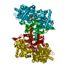
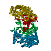
- PDB-1ygp: PHOSPHORYLATED FORM OF YEAST GLYCOGEN PHOSPHORYLASE WITH PHOSPHAT... -
+ Open data

- Basic information
| Entry | Database: PDB / ID: 1ygp | ||||||
|---|---|---|---|---|---|---|---|
| Title | PHOSPHORYLATED FORM OF YEAST GLYCOGEN PHOSPHORYLASE WITH PHOSPHATE BOUND IN THE ACTIVE SITE. | ||||||
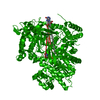
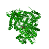
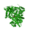
 Components Components | YEAST GLYCOGEN PHOSPHORYLASE | ||||||
 Keywords Keywords | GLYCOSYLTRANSFERASE / YEAST / PHOSPHORYLATED FORM | ||||||
| Function / homology |  Function and homology information Function and homology informationGlycogen breakdown (glycogenolysis) / glycogen phosphorylase / glycogen phosphorylase activity / glycogen catabolic process / Neutrophil degranulation / pyridoxal phosphate binding / cytosol / cytoplasm Similarity search - Function | ||||||
| Biological species |  | ||||||
| Method |  X-RAY DIFFRACTION / Resolution: 2.8 Å X-RAY DIFFRACTION / Resolution: 2.8 Å | ||||||
 Authors Authors | Lin, K. / Rath, V.L. / Dai, S.C. / Fletterick, R.J. / Hwang, P.K. | ||||||
 Citation Citation | Journal: Science / Year: 1996 Title: A protein phosphorylation switch at the conserved allosteric site in GP. Authors: Lin, K. / Rath, V.L. / Dai, S.C. / Fletterick, R.J. / Hwang, P.K. #1: Journal: J.Biol.Chem. / Year: 1995Title: Mechanism of Regulation in Yeast Glycogen Phosphorylase Authors: Lin, K. / Hwang, P.K. / Fletterick, R.J. #2: Journal: Nat.Struct.Biol. / Year: 1994Title: Parallel Evolution in Two Homologues of Phosphorylase Authors: Rath, V.L. / Fletterick, R.J. #3: Journal: J.Mol.Biol. / Year: 1992Title: Purification and Crystallization of Glycogen Phosphorylase from Saccharomyces Cerevisiae Authors: Rath, V.L. / Hwang, P.K. / Fletterick, R.J. | ||||||
| History |
|
- Structure visualization
| Structure viewer | Molecule: MolmilJmol/JSmol |
|---|
- Downloads & links
-Download
| PDBx/mmCIF format | 1ygp.cif.gz | 352.6 KB | Display | PDBx/mmCIF format |
|---|---|---|---|---|
| PDB format | pdb1ygp.ent.gz | 277.6 KB | Display | PDB format |
| PDBx/mmJSON format | 1ygp.json.gz | Tree view | PDBx/mmJSON format | |
| Others |  Other downloads Other downloads |
-Validation report
| Arichive directory | https://data.pdbj.org/pub/pdb/validation_reports/yg/1ygpftp://data.pdbj.org/pub/pdb/validation_reports/yg/1ygp | HTTPS FTP |
|---|
-Related structure data
| Similar structure data |
|---|

-Links
 PDBj
PDBj


- Assembly
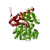
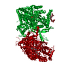
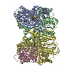
| Deposited unit | 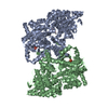
| ||||||||
|---|---|---|---|---|---|---|---|---|---|
| 1 |
| ||||||||
| Unit cell |
| ||||||||
| Noncrystallographic symmetry (NCS) | NCS oper: (Code: given Matrix: (0.756, 0.22, -0.617), Vector: Details | THE AUTHORS ONLY DEPOSITED ONE CHAIN. THE PDB GENERATED THE SECOND CHAIN IN THE ASYMMETRIC UNIT FROM THE CHAIN THAT WAS DEPOSITED USING THE TRANSFORMATION ON MTRIX RECORDS BELOW. | |
-Components
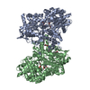
| #1: Protein | Mass: 101278.820 Da / Num. of mol.: 2 / Mutation: N-TERMINAL 22 RESIDUE DELETION Source method: isolated from a genetically manipulated source Details: PHOSPHORYLATED FORM OF THE ENZYME Source: (gene. exp.) Gene: YEAST GLYCOGEN PHOSPHORYLASE / Organ: TAIL / Plasmid: PTACTAC / Gene (production host): YEAST GLYCOGEN PHOSPHORYLASE / Production host:  #2: Chemical | ChemComp-PO4 /   Mass: 94.971 Da / Num. of mol.: 4 / Source method: obtained synthetically / Formula: PO4 Mass: 94.971 Da / Num. of mol.: 4 / Source method: obtained synthetically / Formula: PO4#3: Chemical |   Mass: 247.142 Da / Num. of mol.: 2 / Source method: obtained synthetically / Formula: C8H10NO6P Mass: 247.142 Da / Num. of mol.: 2 / Source method: obtained synthetically / Formula: C8H10NO6PSequence details | THE NUMBERING OF RESIDUES IS RELATIVE TO THE MAMMALIAN GLYCOGEN PHOSPHORYLASE SEQUENCE. THE LONGER ...THE NUMBERING OF RESIDUES IS RELATIVE TO THE MAMMALIAN GLYCOGEN PHOSPHORYL | |
|---|
-Experimental details
-Experiment
| Experiment | Method: X-RAY DIFFRACTION |
|---|
- Sample preparation
| Crystal | Density Matthews: 3.12 Å3/Da / Density % sol: 50 % | ||||||||||||||||||||||||||||||||||||||||||||||||||||||||||||||||||||||||||||||||||||||||||||||||||||||||||||||||
|---|---|---|---|---|---|---|---|---|---|---|---|---|---|---|---|---|---|---|---|---|---|---|---|---|---|---|---|---|---|---|---|---|---|---|---|---|---|---|---|---|---|---|---|---|---|---|---|---|---|---|---|---|---|---|---|---|---|---|---|---|---|---|---|---|---|---|---|---|---|---|---|---|---|---|---|---|---|---|---|---|---|---|---|---|---|---|---|---|---|---|---|---|---|---|---|---|---|---|---|---|---|---|---|---|---|---|---|---|---|---|---|---|---|
| Crystal grow | *PLUS Temperature: 18 ℃ / pH: 6 / Method: vapor diffusion, hanging dropDetails: used to seeding, Lin, K., (1997) Structure, 5, 1511. | ||||||||||||||||||||||||||||||||||||||||||||||||||||||||||||||||||||||||||||||||||||||||||||||||||||||||||||||||
| Components of the solutions | *PLUS
|
-Data collection
| Diffraction source | Wavelength: 1.5418 |
|---|---|
| Detector | Type: RIGAKU RAXIS / Detector: IMAGE PLATE / Date: Apr 8, 1995 |
| Radiation | Monochromatic (M) / Laue (L): M / Scattering type: x-ray |
| Radiation wavelength | Wavelength: 1.5418 Å / Relative weight: 1 |
| Reflection | Num. obs: 50707 / % possible obs: 72 % / Observed criterion σ(I): 1.5 / Redundancy: 5 % / Rmerge(I) obs: 0.105 |
| Reflection | *PLUS Highest resolution: 2.8 Å / Num. measured all: 247322 |
| Reflection shell | *PLUS Highest resolution: 2.8 Å / Lowest resolution: 3 Å / Redundancy: 0.043 % |
- Processing
| Software |
| ||||||||||||||||||||||||||||||||||||||||||||||||||||||||||||
|---|---|---|---|---|---|---|---|---|---|---|---|---|---|---|---|---|---|---|---|---|---|---|---|---|---|---|---|---|---|---|---|---|---|---|---|---|---|---|---|---|---|---|---|---|---|---|---|---|---|---|---|---|---|---|---|---|---|---|---|---|---|
| Refinement | Resolution: 2.8→6 Å / σ(F): 2 Details: RESIDUE PHE 750 IS AT THE BEGINNING OF A BROKEN LOOP FOR WHICH THE ELECTRON DENSITY IS POOR. IT HAS A BAD PHI/PSI ANGLE, AND SHOULD BE IGNORED.
| ||||||||||||||||||||||||||||||||||||||||||||||||||||||||||||
| Refinement step | Cycle: LAST / Resolution: 2.8→6 Å
| ||||||||||||||||||||||||||||||||||||||||||||||||||||||||||||
| Refine LS restraints |
| ||||||||||||||||||||||||||||||||||||||||||||||||||||||||||||
| Xplor file |
|
