 Movie
Movie Controller

[English] 日本語
 Yorodumi
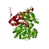
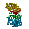
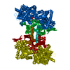
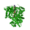
- PDB-3mt8: Glycogen phosphorylase complexed with 4-chlorobenzaldehyde-4-(bet... -
+ Open data

- Basic information
| Entry | Database: PDB / ID: 3mt8 | ||||||
|---|---|---|---|---|---|---|---|
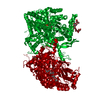
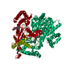
| Title | Glycogen phosphorylase complexed with 4-chlorobenzaldehyde-4-(beta-D-glucopyranosyl)-thiosemicarbazone | ||||||
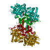
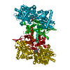
 Components Components | Glycogen phosphorylase, muscle form | ||||||
 Keywords Keywords | TRANSFERASE/TRANSFERASE INHIBITOR / Glycogenolysis / type 2 diabetes / TRANSFERASE-TRANSFERASE INHIBITOR complex | ||||||
| Function / homology |  Function and homology information Function and homology informationglycogen phosphorylase / glycogen phosphorylase activity / glycogen catabolic process / skeletal muscle myofibril / pyridoxal phosphate binding / nucleotide binding Similarity search - Function | ||||||
| Biological species |  | ||||||
| Method |  X-RAY DIFFRACTION / SYNCHROTRON / FOURIER SYNTHESIS / Resolution: 2 Å X-RAY DIFFRACTION / SYNCHROTRON / FOURIER SYNTHESIS / Resolution: 2 Å | ||||||
 Authors Authors | Alexacou, K.-M. | ||||||
 Citation Citation | Journal: Bioorg.Med.Chem. / Year: 2010 Title: The binding of beta-D-glucopyranosyl-thiosemicarbazone derivatives to glycogen phosphorylase: a new class of inhibitors Authors: Alexacou, K.M. / Tenchiu Deleanu, A.C. / Chrysina, E.D. / Charavgi, M.D. / Kostas, I.D. / Zographos, S.E. / Oikonomakos, N.G. / Leonidas, D.D. | ||||||
| History |
|
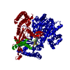
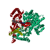
- Structure visualization
| Structure viewer | Molecule: MolmilJmol/JSmol |
|---|
- Downloads & links
-Download
| PDBx/mmCIF format | 3mt8.cif.gz | 182.4 KB | Display | PDBx/mmCIF format |
|---|---|---|---|---|
| PDB format | pdb3mt8.ent.gz | 142.2 KB | Display | PDB format |
| PDBx/mmJSON format | 3mt8.json.gz | Tree view | PDBx/mmJSON format | |
| Others |  Other downloads Other downloads |
-Validation report
| Arichive directory | https://data.pdbj.org/pub/pdb/validation_reports/mt/3mt8ftp://data.pdbj.org/pub/pdb/validation_reports/mt/3mt8 | HTTPS FTP |
|---|
-Related structure data
| Related structure data | 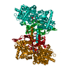 3mqfC  3mrtC 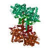 3mrvC  3mrxC 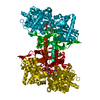 3ms2C  3ms4C 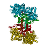 3ms7C  3mscC  3mt7C 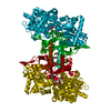 3mt9C 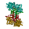 3mtaC  3mtbC 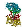 3mtdC  3nc4C 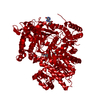 2prjS C: citing same article ( S: Starting model for refinement |
|---|---|
| Similar structure data |
-Links
 PDBj
PDBj
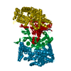
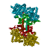
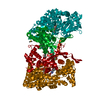
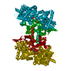
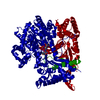
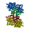
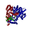
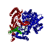
- Assembly
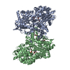
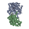
| Deposited unit | 
| ||||||||
|---|---|---|---|---|---|---|---|---|---|
| 1 | 
| ||||||||
| Unit cell |
|
-Components
| #1: Protein | Mass: 97519.320 Da / Num. of mol.: 1 / Source method: isolated from a natural source / Details: Muscle / Source: (natural) | ||
|---|---|---|---|
| #2: Sugar |   Type: D-saccharide / Mass: 375.828 Da / Num. of mol.: 2 / Source method: obtained synthetically / Formula: C14H18ClN3O5S Type: D-saccharide / Mass: 375.828 Da / Num. of mol.: 2 / Source method: obtained synthetically / Formula: C14H18ClN3O5S#3: Water | ChemComp-HOH / |  Mass: 18.015 Da / Num. of mol.: 292 / Source method: isolated from a natural source / Formula: H2O Mass: 18.015 Da / Num. of mol.: 292 / Source method: isolated from a natural source / Formula: H2O |
-Experimental details
-Experiment
| Experiment | Method: X-RAY DIFFRACTION / Number of used crystals: 1 |
|---|
- Sample preparation
| Crystal | Density Matthews: 2.44 Å3/Da / Density % sol: 49.61 % |
|---|---|
| Crystal grow | Temperature: 289 K / Method: small tubes / pH: 6.7 Details: Crystals grown from 20 mg/ml protein in a buffer of 10 mM BES, pH 6.7, 1mM EDTA, 3mM DTT. Crystals soaked with 20mM inhibitor in 20% DMSO for 21 hrs, SMALL TUBES, temperature 289K |
-Data collection
| Diffraction | Mean temperature: 293 K |
|---|---|
| Diffraction source | Source: SYNCHROTRON / Site: SRS  / Beamline: PX10.1 / Wavelength: 1.04498 Å / Beamline: PX10.1 / Wavelength: 1.04498 Å |
| Detector | Type: MARMOSAIC 225 mm CCD / Detector: CCD / Date: Nov 19, 2007 |
| Radiation | Monochromator: Crystal / Protocol: SINGLE WAVELENGTH / Monochromatic (M) / Laue (L): M / Scattering type: x-ray |
| Radiation wavelength | Wavelength: 1.04498 Å / Relative weight: 1 |
| Reflection | Resolution: 2→35.69 Å / Num. all: 63139 / Num. obs: 63139 / % possible obs: 97 % / Observed criterion σ(F): 0 / Observed criterion σ(I): -3 / Redundancy: 5.4 % / Biso Wilson estimate: 27.4 Å2 / Rmerge(I) obs: 0.054 / Net I/σ(I): 21.3 |
| Reflection shell | Resolution: 2→2.11 Å / Redundancy: 5.4 % / Rmerge(I) obs: 0.221 / Mean I/σ(I) obs: 7.2 / Num. unique all: 9172 / % possible all: 97.1 |
- Processing
| Software |
| ||||||||||||||||||||||||||||||||||||||||||||||||||||||||||||||||||||||||||||||||||||||||||
|---|---|---|---|---|---|---|---|---|---|---|---|---|---|---|---|---|---|---|---|---|---|---|---|---|---|---|---|---|---|---|---|---|---|---|---|---|---|---|---|---|---|---|---|---|---|---|---|---|---|---|---|---|---|---|---|---|---|---|---|---|---|---|---|---|---|---|---|---|---|---|---|---|---|---|---|---|---|---|---|---|---|---|---|---|---|---|---|---|---|---|---|
| Refinement | Method to determine structure: FOURIER SYNTHESIS Starting model: 2PRJ Resolution: 2→35.69 Å / Cor.coef. Fo:Fc: 0.957 / Cor.coef. Fo:Fc free: 0.946 / SU B: 3.61 / SU ML: 0.102 / Isotropic thermal model: RESTRAINED / Cross valid method: THROUGHOUT / ESU R: 0.186 / ESU R Free: 0.15 / Stereochemistry target values: MAXIMUM LIKELIHOOD / Details: HYDROGENS HAVE BEEN ADDED IN THE RIDING POSITIONS
| ||||||||||||||||||||||||||||||||||||||||||||||||||||||||||||||||||||||||||||||||||||||||||
| Solvent computation | Ion probe radii: 0.8 Å / Shrinkage radii: 0.8 Å / VDW probe radii: 1.2 Å / Solvent model: MASK | ||||||||||||||||||||||||||||||||||||||||||||||||||||||||||||||||||||||||||||||||||||||||||
| Displacement parameters | Biso mean: 31.102 Å2
| ||||||||||||||||||||||||||||||||||||||||||||||||||||||||||||||||||||||||||||||||||||||||||
| Refinement step | Cycle: LAST / Resolution: 2→35.69 Å
| ||||||||||||||||||||||||||||||||||||||||||||||||||||||||||||||||||||||||||||||||||||||||||
| Refine LS restraints |
| ||||||||||||||||||||||||||||||||||||||||||||||||||||||||||||||||||||||||||||||||||||||||||
| LS refinement shell | Resolution: 2→2.052 Å / Total num. of bins used: 20
|
