 Movie
Movie Controller

[English] 日本語
 Yorodumi
- PDB-1fu7: STRUCTURES OF GLYCOGEN PHOSPHORYLASE-INHIBITOR COMPLEXES AND THE ... -
+ Open data

- Basic information
| Entry | Database: PDB / ID: 1fu7 | ||||||
|---|---|---|---|---|---|---|---|
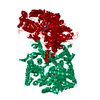
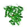
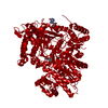
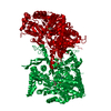
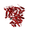
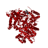
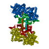
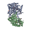
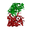
| Title | STRUCTURES OF GLYCOGEN PHOSPHORYLASE-INHIBITOR COMPLEXES AND THE IMPLICATIONS FOR STRUCTURE-BASED DRUG DESIGN | ||||||
 Components Components | GLYCOGEN PHOSPHORYLASE | ||||||
 Keywords Keywords | TRANSFERASE / GLYCOGEN PHOSPHORYLASE / INHIBITOR COMPLEX / CATALYTIC SITE / DESIGN | ||||||
| Function / homology |  Function and homology information Function and homology informationglycogen phosphorylase / glycogen phosphorylase activity / glycogen catabolic process / skeletal muscle myofibril / pyridoxal phosphate binding / nucleotide binding Similarity search - Function | ||||||
| Biological species |  | ||||||
| Method |  X-RAY DIFFRACTION / OTHER / Resolution: 2.36 Å X-RAY DIFFRACTION / OTHER / Resolution: 2.36 Å | ||||||
 Authors Authors | Watson, K.A. / Tsitsanou, K.E. / Gregoriou, M. / Zographos, S.E. / Skamnaki, V.T. / Oikonomakos, N.G. / Fleet, G.W. / Johnson, L.N. | ||||||
 Citation Citation | Journal: Proteins / Year: 2005 Title: Kinetic and crystallographic studies of glucopyranose spirohydantoin and glucopyranosylamine analogs inhibitors of glycogen phosphorylase. Authors: Watson, K.A. / Chrysina, E.D. / Tsitsanou, K.E. / Zographos, S.E. / Archontis, G. / Fleet, G.W. / Oikonomakos, N.G. #1: Journal: Protein Sci. / Year: 1998Title: The Structure of a Glycogen Phosphorylase Glucopyranose Spirohydantoin Complex at 1.8 A Resolution and 100K: The Role of the Water Structure and its Contribution to Binding Authors: Gregoriou, M. / Noble, M.E. / Watson, K.A. / Garman, E.F. / Krulle, T.M. / de la Fuente, C. / Fleet, G.W. / Oikonomakos, N.G. / Johnson, L.N. #2: Journal: Tetrahedron Lett. / Year: 1995Title: Potent Inhibition of Glycogen Phosphorylase by a Spirohydantoin of Glucopyranose: First Pyranose Analogues of Hydantocidin Authors: Bichard, C.J.F. / Mitchell, E.P. / Wormald, M.R. / Watson, K.A. / Johnson, L.N. / Zographos, S.E. / Koutra, D.D. / Oikonomakos, N.G. / Fleet, G.W.J. #3: Journal: Protein Sci. / Year: 1995Title: N-Acetyl-Beta-D-Glucopyranosylamine: A Potent T-State Inhibitor of Glycogen Phosphorylase. A Comparison with Alpha-D-Glucose Authors: Oikonomakos, N.G. / Kontou, M. / Zographos, S.E. / Watson, K.A. / Johnson, L.N. / Bichard, C.J. / Fleet, G.W. / Acharya, K.R. #4: Journal: Biochemistry / Year: 1994Title: Design of Inhibitors of Glycogen Phosphorylase: A Study of Alpha- and Beta-C-Glucosides and 1-Thio-Beta-D-Glucose Compounds Authors: Watson, K.A. / Mitchell, E.P. / Johnson, L.N. / Son, J.C. / Bichard, C.J. / Orchard, M.G. / Fleet, G.W. / Oikonomakos, N.G. / Leonidas, D.D. / Kontou, M. / Papageorgiou, A.C. | ||||||
| History |
|
- Structure visualization
| Structure viewer | Molecule: MolmilJmol/JSmol |
|---|
- Downloads & links
-Download
| PDBx/mmCIF format | 1fu7.cif.gz | 183.9 KB | Display | PDBx/mmCIF format |
|---|---|---|---|---|
| PDB format | pdb1fu7.ent.gz | 145.1 KB | Display | PDB format |
| PDBx/mmJSON format | 1fu7.json.gz | Tree view | PDBx/mmJSON format | |
| Others |  Other downloads Other downloads |
-Validation report
| Arichive directory | https://data.pdbj.org/pub/pdb/validation_reports/fu/1fu7ftp://data.pdbj.org/pub/pdb/validation_reports/fu/1fu7 | HTTPS FTP |
|---|
-Related structure data
| Related structure data |  1fs4C  1ftqC  1ftwC  1ftyC  1fu4C  1fu8C  2prjS C: citing same article ( S: Starting model for refinement |
|---|---|
| Similar structure data |


-Links
 PDBj
PDBj
- Assembly
| Deposited unit | 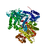
| ||||||||
|---|---|---|---|---|---|---|---|---|---|
| 1 | 
| ||||||||
| Unit cell |
|
-Components
| #1: Protein | Mass: 97291.203 Da / Num. of mol.: 1 / Source method: isolated from a natural source / Source: (natural) |
|---|---|
| #2: Sugar | ChemComp-CR1 /   Type: D-saccharide / Mass: 237.207 Da / Num. of mol.: 1 Type: D-saccharide / Mass: 237.207 Da / Num. of mol.: 1Source method: isolated from a genetically manipulated source Formula: C8H15NO7 |
| #3: Chemical | ChemComp-PLP /   Mass: 247.142 Da / Num. of mol.: 1 / Source method: obtained synthetically / Formula: C8H10NO6P Mass: 247.142 Da / Num. of mol.: 1 / Source method: obtained synthetically / Formula: C8H10NO6P |
| #4: Water | ChemComp-HOH /  Mass: 18.015 Da / Num. of mol.: 218 / Source method: isolated from a natural source / Formula: H2O Mass: 18.015 Da / Num. of mol.: 218 / Source method: isolated from a natural source / Formula: H2O |
-Experimental details
-Experiment
| Experiment | Method: X-RAY DIFFRACTION / Number of used crystals: 1 |
|---|
- Sample preparation
| Crystal | Density Matthews: 2.47 Å3/Da / Density % sol: 48 % | |||||||||||||||||||||||||||||||||||
|---|---|---|---|---|---|---|---|---|---|---|---|---|---|---|---|---|---|---|---|---|---|---|---|---|---|---|---|---|---|---|---|---|---|---|---|---|
| Crystal grow | Temperature: 289 K / Method: small tubes / pH: 6.7 Details: 10 mM BES, 0.1 MM EDTA, 1 mM IMP, 1 mM spermine , pH 6.70, SMALL TUBES, temperature 289K | |||||||||||||||||||||||||||||||||||
| Crystal grow | *PLUS Temperature: 16 ℃ / pH: 6.7 / Method: unknownDetails: Oikonomakos, N.G., (1985) Biochim.Biophys.Acta., 832, 248. | |||||||||||||||||||||||||||||||||||
| Components of the solutions | *PLUS
|
-Data collection
| Diffraction | Mean temperature: 298 K |
|---|---|
| Diffraction source | Type: OTHER / Wavelength: 1.5418 |
| Detector | Type: NICOLET IPC / Detector: AREA DETECTOR / Date: Jan 18, 1996 |
| Radiation | Monochromator: NI FILTER / Protocol: SINGLE WAVELENGTH / Monochromatic (M) / Laue (L): M / Scattering type: x-ray |
| Radiation wavelength | Wavelength: 1.5418 Å / Relative weight: 1 |
| Reflection | Resolution: 2.36→14.8 Å / Num. obs: 32936 / % possible obs: 82 % / Rmerge(I) obs: 0.062 |
| Reflection shell | Resolution: 2.36→2.47 Å |
| Reflection | *PLUS Highest resolution: 2.35 Å / Lowest resolution: 14.8 Å / % possible obs: 82 % / Num. measured all: 52973 |
- Processing
| Software |
| ||||||||||||||||||||||||||||||||||||||||||||||||||||||||||||
|---|---|---|---|---|---|---|---|---|---|---|---|---|---|---|---|---|---|---|---|---|---|---|---|---|---|---|---|---|---|---|---|---|---|---|---|---|---|---|---|---|---|---|---|---|---|---|---|---|---|---|---|---|---|---|---|---|---|---|---|---|---|
| Refinement | Method to determine structure: OTHER Starting model: 2PRJ Resolution: 2.36→14.8 Å / Cross valid method: THROUGHOUT / σ(F): 0 / Stereochemistry target values: Engh & Huber
| ||||||||||||||||||||||||||||||||||||||||||||||||||||||||||||
| Refinement step | Cycle: LAST / Resolution: 2.36→14.8 Å
| ||||||||||||||||||||||||||||||||||||||||||||||||||||||||||||
| Refine LS restraints |
| ||||||||||||||||||||||||||||||||||||||||||||||||||||||||||||
| Software | *PLUS Name: X-PLOR / Version: 3.851 / Classification: refinement | ||||||||||||||||||||||||||||||||||||||||||||||||||||||||||||
| Refinement | *PLUS Lowest resolution: 14.8 Å / % reflection Rfree: 5 % / Rfactor obs: 0.168 | ||||||||||||||||||||||||||||||||||||||||||||||||||||||||||||
| Solvent computation | *PLUS | ||||||||||||||||||||||||||||||||||||||||||||||||||||||||||||
| Displacement parameters | *PLUS | ||||||||||||||||||||||||||||||||||||||||||||||||||||||||||||
| Refine LS restraints | *PLUS Type: x_angle_deg / Dev ideal: 1.3 |
