 Movie
Movie Controller

[English] 日本語
 Yorodumi
- PDB-1c50: IDENTIFICATION AND STRUCTURAL CHARACTERIZATION OF A NOVEL ALLOSTE... -
+ Open data

- Basic information
| Entry | Database: PDB / ID: 1c50 | ||||||
|---|---|---|---|---|---|---|---|
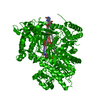
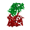
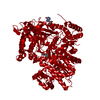
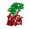
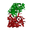
| Title | IDENTIFICATION AND STRUCTURAL CHARACTERIZATION OF A NOVEL ALLOSTERIC BINDING SITE OF GLYCOGEN PHOSPHORYLASE B | ||||||
 Components Components | PROTEIN (GLYCOGEN PHOSPHORYLASE) | ||||||
 Keywords Keywords | TRANSFERASE / GLYCOGEN METABOLISM / GLYCOGEN PHOSPHORYLASE B / INHIBITION / CENTRAL CAVITY / DRUG BINDING SITE | ||||||
| Function / homology |  Function and homology information Function and homology informationglycogen phosphorylase / glycogen phosphorylase activity / glycogen catabolic process / skeletal muscle myofibril / pyridoxal phosphate binding / nucleotide binding Similarity search - Function | ||||||
| Biological species |  | ||||||
| Method |  X-RAY DIFFRACTION / OTHER / Resolution: 2.3 Å X-RAY DIFFRACTION / OTHER / Resolution: 2.3 Å | ||||||
 Authors Authors | Oikonomakos, N.G. / Skamnaki, V.T. / Tsitsanou, K.E. / Gavalas, N.G. / Johnson, L.N. | ||||||
 Citation Citation | Journal: Structure Fold.Des. / Year: 2000 Title: A new allosteric site in glycogen phosphorylase b as a target for drug interactions. Authors: Oikonomakos, N.G. / Skamnaki, V.T. / Tsitsanou, K.E. / Gavalas, N.G. / Johnson, L.N. #1: Journal: Protein Sci. / Year: 1999Title: Allosteric Inhibition of Glycogen Phosphorylase a by the Potential Antidiabetic Drug 3-Isopropyl 4-(2-Chlorophenyl)-1,4-Dihydro-1-Ethyl-2-Methyl-Pyridine-3,5,6-Tricarboxylate Authors: Oikonomakos, N.G. / Tsitsanou, K.E. / Zographos, S.E. / Skamnaki, V.T. / Goldmann, S. / Bischoff, H. #2: Journal: Structure / Year: 1997Title: The Structure of Glycogen Phosphorylase b with an Alkyldihydropyridine-Dicarboxylic Acid Compound, A Novel and Potent Inhibitor Authors: Zographos, S.E. / Oikonomakos, N.G. / Tsitsanou, K.E. / Leonidas, D.D. / Chrysina, E.D. / Skamnaki, V.T. / Bischoff, H. / Goldmann, S. / Watson, K.A. / Johnson, L.N. | ||||||
| History |
|
- Structure visualization
| Structure viewer | Molecule: MolmilJmol/JSmol |
|---|
- Downloads & links
-Download
| PDBx/mmCIF format | 1c50.cif.gz | 186.4 KB | Display | PDBx/mmCIF format |
|---|---|---|---|---|
| PDB format | pdb1c50.ent.gz | 145.3 KB | Display | PDB format |
| PDBx/mmJSON format | 1c50.json.gz | Tree view | PDBx/mmJSON format | |
| Others |  Other downloads Other downloads |
-Validation report
| Arichive directory | https://data.pdbj.org/pub/pdb/validation_reports/c5/1c50ftp://data.pdbj.org/pub/pdb/validation_reports/c5/1c50 | HTTPS FTP |
|---|
-Related structure data
| Similar structure data |
|---|


-Links
 PDBj
PDBj
- Assembly
| Deposited unit | 
| ||||||||
|---|---|---|---|---|---|---|---|---|---|
| 1 | 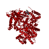
| ||||||||
| Unit cell |
|
-Components
| #1: Protein | Mass: 95833.562 Da / Num. of mol.: 1 / Source method: isolated from a natural source / Source: (natural) |
|---|---|
| #2: Chemical | ChemComp-PLP /   Mass: 247.142 Da / Num. of mol.: 1 / Source method: obtained synthetically / Formula: C8H10NO6P Mass: 247.142 Da / Num. of mol.: 1 / Source method: obtained synthetically / Formula: C8H10NO6P |
| #3: Chemical | ChemComp-CHI /   Mass: 443.898 Da / Num. of mol.: 1 / Source method: obtained synthetically / Formula: C23H23ClFN3O3 Mass: 443.898 Da / Num. of mol.: 1 / Source method: obtained synthetically / Formula: C23H23ClFN3O3 |
| #4: Water | ChemComp-HOH /  Mass: 18.015 Da / Num. of mol.: 256 / Source method: isolated from a natural source / Formula: H2O Mass: 18.015 Da / Num. of mol.: 256 / Source method: isolated from a natural source / Formula: H2O |
| Sequence details | REFERENCE: K.NAKANO, P.K.HWANG, R.J.FLETTERICK, FEBS LETT., V. 204, P. 283, 1986. HIGH RESOLUTION ...REFERENCE: K.NAKANO, P.K.HWANG, R.J.FLETTERICK |
-Experimental details
-Experiment
| Experiment | Method: X-RAY DIFFRACTION / Number of used crystals: 1 |
|---|
- Sample preparation
| Crystal | Density Matthews: 2.49 Å3/Da / Density % sol: 48 % | ||||||||||||||||||||||||||||||||||||||||
|---|---|---|---|---|---|---|---|---|---|---|---|---|---|---|---|---|---|---|---|---|---|---|---|---|---|---|---|---|---|---|---|---|---|---|---|---|---|---|---|---|---|
| Crystal grow | pH: 6.7 Details: PHOSPHORYLASE B-CP320626 COMPLEX WAS CO-CRYSTALLISED UNDER CONDITIONS SIMILAR TO THOSE DESCRIBED BY ZOGRAPHOS ET AL., (1997) STRUCTURE 5, 1413-1425., pH 6.7 | ||||||||||||||||||||||||||||||||||||||||
| Crystal grow | *PLUS Temperature: 16 ℃ / Method: unknownDetails: Zographos, S.E., (1997) Structure (London), 5, 1413. | ||||||||||||||||||||||||||||||||||||||||
| Components of the solutions | *PLUS
|
-Data collection
| Diffraction | Mean temperature: 287 K |
|---|---|
| Diffraction source | Source: ROTATING ANODE / Wavelength: 1.5418 |
| Detector | Type: MAR scanner 180 mm plate / Detector: IMAGE PLATE / Date: Jul 15, 1999 |
| Radiation | Protocol: SINGLE WAVELENGTH / Monochromatic (M) / Laue (L): M / Scattering type: x-ray |
| Radiation wavelength | Wavelength: 1.5418 Å / Relative weight: 1 |
| Reflection | Resolution: 2.3→30 Å / Num. obs: 42617 / % possible obs: 96.3 % / Observed criterion σ(I): 0 / Redundancy: 3.8 % / Rmerge(I) obs: 0.11 / Net I/σ(I): 6.8 |
| Reflection shell | Resolution: 2.3→2.34 Å / Rmerge(I) obs: 0.445 / Mean I/σ(I) obs: 1.7 / % possible all: 91 |
| Reflection | *PLUS Num. measured all: 160475 / Rmerge(I) obs: 0.11 |
| Reflection shell | *PLUS % possible obs: 91 % |
- Processing
| Software |
| ||||||||||||||||||||||||||||||||||||||||||||||||||||||||||||
|---|---|---|---|---|---|---|---|---|---|---|---|---|---|---|---|---|---|---|---|---|---|---|---|---|---|---|---|---|---|---|---|---|---|---|---|---|---|---|---|---|---|---|---|---|---|---|---|---|---|---|---|---|---|---|---|---|---|---|---|---|---|
| Refinement | Method to determine structure: OTHER / Resolution: 2.3→30 Å / Cross valid method: R FREE / σ(F): 0
| ||||||||||||||||||||||||||||||||||||||||||||||||||||||||||||
| Refinement step | Cycle: LAST / Resolution: 2.3→30 Å
| ||||||||||||||||||||||||||||||||||||||||||||||||||||||||||||
| Refine LS restraints |
| ||||||||||||||||||||||||||||||||||||||||||||||||||||||||||||
| Software | *PLUS Name: X-PLOR / Version: 3.8 / Classification: refinement | ||||||||||||||||||||||||||||||||||||||||||||||||||||||||||||
| Refinement | *PLUS Highest resolution: 2.3 Å / Lowest resolution: 30 Å / σ(F): 0 / Num. reflection Rfree: 2162 / % reflection Rfree: 5 % | ||||||||||||||||||||||||||||||||||||||||||||||||||||||||||||
| Solvent computation | *PLUS | ||||||||||||||||||||||||||||||||||||||||||||||||||||||||||||
| Displacement parameters | *PLUS | ||||||||||||||||||||||||||||||||||||||||||||||||||||||||||||
| Refine LS restraints | *PLUS
|
