- PDB-1axr: COOPERATIVITY BETWEEN HYDROGEN-BONDING AND CHARGE-DIPOLE INTERACT... -
+
Open data
ID or keywords:
Loading...
-
Basic information
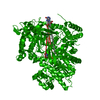
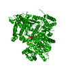
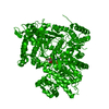
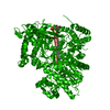
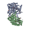
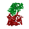
Entry
Database: PDB / ID: 1axr
Title
COOPERATIVITY BETWEEN HYDROGEN-BONDING AND CHARGE-DIPOLE INTERACTIONS IN THE INHIBITION OF BETA-GLYCOSIDASES BY AZOLOPYRIDINES: EVIDENCE FROM A STUDY WITH GLYCOGEN PHOSPHORYLASE B
Journal: HELV.CHIM.ACTA / Year: 1998 Title: Cooperative interactions of the catalytic nucleophile and the catalytic acid in the inhibition of beta-glycosidases. Calculations and their validation by comparative kinetic and structural ...Title: Cooperative interactions of the catalytic nucleophile and the catalytic acid in the inhibition of beta-glycosidases. Calculations and their validation by comparative kinetic and structural studies of the inhibition of glycogen phosphorylase b. Authors: Heightman, T.D. / Vasella, A. / Tsitsanou, K.E. / Zographos, S.E. / Skamnaki, V.T. / Oikonomakos, N.G.
Mass: 18.015 Da / Num. of mol.: 500 / Source method: isolated from a natural source / Formula: H2O
-
Experimental details
-
Experiment
Experiment
Method: X-RAY DIFFRACTION / Number of used crystals: 1
-
Sample preparation
Crystal
Density Matthews: 2.38 Å3/Da / Density % sol: 48 %
Crystal grow
Method: soak native crystals / pH: 6.7 Details: NATIVE T-STATE GLYCOGEN PHOSPHORYLASE CRYSTALS WERE SOAKED WITH 10 MM BES, 0.1 MM EDTA AND 0.02% SODIUM AZIDE SOLUTION CONTAINING 5 MM TRIAZOLE AND 50 MM SODIUM DIHYDROGEN PHOSPHATE FOR 1 ...Details: NATIVE T-STATE GLYCOGEN PHOSPHORYLASE CRYSTALS WERE SOAKED WITH 10 MM BES, 0.1 MM EDTA AND 0.02% SODIUM AZIDE SOLUTION CONTAINING 5 MM TRIAZOLE AND 50 MM SODIUM DIHYDROGEN PHOSPHATE FOR 1 HOUR., pH 6.7, soak native crystals
In the structure databanks used in Yorodumi, some data are registered as the other names, "COVID-19 virus" and "2019-nCoV". Here are the details of the virus and the list of structure data.
Jan 31, 2019. EMDB accession codes are about to change! (news from PDBe EMDB page)
EMDB accession codes are about to change! (news from PDBe EMDB page)
The allocation of 4 digits for EMDB accession codes will soon come to an end. Whilst these codes will remain in use, new EMDB accession codes will include an additional digit and will expand incrementally as the available range of codes is exhausted. The current 4-digit format prefixed with “EMD-” (i.e. EMD-XXXX) will advance to a 5-digit format (i.e. EMD-XXXXX), and so on. It is currently estimated that the 4-digit codes will be depleted around Spring 2019, at which point the 5-digit format will come into force.
The EM Navigator/Yorodumi systems omit the EMD- prefix.
Related info.:Q: What is EMD? / ID/Accession-code notation in Yorodumi/EM Navigator
Yorodumi is a browser for structure data from EMDB, PDB, SASBDB, etc.
This page is also the successor to EM Navigator detail page, and also detail information page/front-end page for Omokage search.
The word "yorodu" (or yorozu) is an old Japanese word meaning "ten thousand". "mi" (miru) is to see.
Related info.:EMDB / PDB / SASBDB / Comparison of 3 databanks / Yorodumi Search / Aug 31, 2016. New EM Navigator & Yorodumi / Yorodumi Papers / Jmol/JSmol / Function and homology information / Changes in new EM Navigator and Yorodumi
 Movie
Movie Controller

 Yorodumi
 Open data
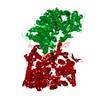
 Basic information
 Components
 Keywords
 Function and homology information
 X-RAY DIFFRACTION /
  Authors
 Citation
 Structure visualization
 Downloads & links
 Other downloads
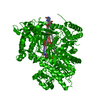


 PDBj
PDBj
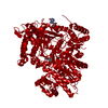
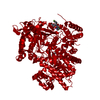
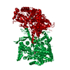
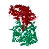
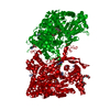
 Assembly
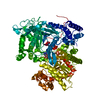

 Mass: 94.971 Da / Num. of mol.: 2 / Source method: obtained synthetically / Formula: PO4
 Mass: 247.142 Da / Num. of mol.: 1 / Source method: obtained synthetically / Formula: C8H10NO6P
 Mass: 202.188 Da / Num. of mol.: 1 / Source method: obtained synthetically / Formula: C7H12N3O4
 Mass: 18.015 Da / Num. of mol.: 500 / Source method: isolated from a natural source / Formula: H2O
 Sample preparation
 / Beamline: 5.2R / Wavelength: 1
  Processing