 Movie
Movie Controller

[English] 日本語
 Yorodumi
- PDB-2amv: THE STRUCTURE OF GLYCOGEN PHOSPHORYLASE B WITH AN ALKYL-DIHYDROPY... -
+ Open data

- Basic information
| Entry | Database: PDB / ID: 2amv | |||||||||
|---|---|---|---|---|---|---|---|---|---|---|
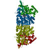
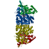
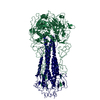
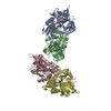
| Title | THE STRUCTURE OF GLYCOGEN PHOSPHORYLASE B WITH AN ALKYL-DIHYDROPYRIDINE-DICARBOXYLIC ACID | |||||||||
 Components Components | PROTEIN (GLYCOGEN PHOSPHORYLASE) | |||||||||
 Keywords Keywords | TRANSFERASE / GLYCOGEN PHOSPHORYLASE / GLYCOGEN METABOLISM / DIABETES / INHIBITORS / GLYCOSYLTRANSFERASE | |||||||||
| Function / homology |  Function and homology information Function and homology informationglycogen phosphorylase / glycogen phosphorylase activity / glycogen catabolic process / skeletal muscle myofibril / pyridoxal phosphate binding / nucleotide binding Similarity search - Function | |||||||||
| Biological species |  | |||||||||
| Method |  X-RAY DIFFRACTION / SYNCHROTRON / OTHER / Resolution: 2.3 Å X-RAY DIFFRACTION / SYNCHROTRON / OTHER / Resolution: 2.3 Å | |||||||||
 Authors Authors | Zographos, S.E. / Oikonomakos, N.G. / Johnson, L.N. | |||||||||
 Citation Citation | Journal: Structure / Year: 1997 Title: The structure of glycogen phosphorylase b with an alkyldihydropyridine-dicarboxylic acid compound, a novel and potent inhibitor. Authors: Zographos, S.E. / Oikonomakos, N.G. / Tsitsanou, K.E. / Leonidas, D.D. / Chrysina, E.D. / Skamnaki, V.T. / Bischoff, H. / Goldmann, S. / Watson, K.A. / Johnson, L.N. #1: Journal: Protein Sci. / Year: 1999Title: Effects of Commonly Used Cryoprotectants on Glycogen Phosphorylase Activity and Structure Authors: Tsitsanou, K.E. / Oikonomakos, N.G. / Zographos, S.E. / Skamnaki, V.T. / Gregoriou, M. / Watson, K.A. / Johnson, L.N. / Fleet, G.W. #2: Journal: Glycogen Phosphorylase B: Description of the Protein StructureYear: 1991 Title: Glycogen Phosphorylase B: Description of the Protein Structure Authors: Acharya, K.R. / Stuart, D.I. / Varvill, K.M. / Johnson, L.N. #3: Journal: J.Mol.Biol. / Year: 1993Title: Crystallographic Binding Studies on the Allosteric Inhibitor Glucose-6- Phosphate to T State Glycogen Phosphorylase b Authors: Johnson, L.N. / Snape, P. / Martin, J.L. / Acharya, K.R. / Barford, D. / Oikonomakos, N.G. | |||||||||
| History |
|
- Structure visualization
| Structure viewer | Molecule: MolmilJmol/JSmol |
|---|
- Downloads & links
-Download
| PDBx/mmCIF format | 2amv.cif.gz | 192.2 KB | Display | PDBx/mmCIF format |
|---|---|---|---|---|
| PDB format | pdb2amv.ent.gz | 151.2 KB | Display | PDB format |
| PDBx/mmJSON format | 2amv.json.gz | Tree view | PDBx/mmJSON format | |
| Others |  Other downloads Other downloads |
-Validation report
| Arichive directory | https://data.pdbj.org/pub/pdb/validation_reports/am/2amvftp://data.pdbj.org/pub/pdb/validation_reports/am/2amv | HTTPS FTP |
|---|
-Related structure data
| Similar structure data |
|---|


-Links
 PDBj
PDBj
- Assembly
| Deposited unit | 
| ||||||||
|---|---|---|---|---|---|---|---|---|---|
| 1 | 
| ||||||||
| 2 |
| ||||||||
| 3 | 
| ||||||||
| Unit cell |
|
-Components
| #1: Protein | Mass: 97291.203 Da / Num. of mol.: 1 / Source method: isolated from a natural source / Source: (natural) |
|---|---|
| #2: Chemical | ChemComp-PLP /   Mass: 247.142 Da / Num. of mol.: 1 / Source method: obtained synthetically / Formula: C8H10NO6P Mass: 247.142 Da / Num. of mol.: 1 / Source method: obtained synthetically / Formula: C8H10NO6P |
| #3: Chemical | ChemComp-BIN /   Mass: 406.837 Da / Num. of mol.: 1 / Source method: obtained synthetically / Formula: C20H21ClNO6 Mass: 406.837 Da / Num. of mol.: 1 / Source method: obtained synthetically / Formula: C20H21ClNO6 |
| #4: Chemical | ChemComp-GOL /   Mass: 92.094 Da / Num. of mol.: 1 / Source method: obtained synthetically / Formula: C3H8O3 Mass: 92.094 Da / Num. of mol.: 1 / Source method: obtained synthetically / Formula: C3H8O3 |
| #5: Water | ChemComp-HOH /  Mass: 18.015 Da / Num. of mol.: 559 / Source method: isolated from a natural source / Formula: H2O Mass: 18.015 Da / Num. of mol.: 559 / Source method: isolated from a natural source / Formula: H2O |
| Has protein modification | N |
-Experimental details
-Experiment
| Experiment | Method: X-RAY DIFFRACTION / Number of used crystals: 1 |
|---|
- Sample preparation
| Crystal | Density Matthews: 2.4 Å3/Da / Density % sol: 48 % | ||||||||||||||||||||||||||||||||||||||||
|---|---|---|---|---|---|---|---|---|---|---|---|---|---|---|---|---|---|---|---|---|---|---|---|---|---|---|---|---|---|---|---|---|---|---|---|---|---|---|---|---|---|
| Crystal grow | Temperature: 289 K / pH: 6.7 Details: PHOSPHORYLASE B WAS COCRYSTALLISED WITH 1 MM W1807 IN A MEDIUM CONSISTING OF 27-28 MG/ML ENZYME, 1 MM SPERMINE, 3 MM DTT, 10 MM BES, 0.1 MM EDTA, AND 0.02% SODIUM AZIDE, PH 6.7 (16 DEG C)., temperature 289K | ||||||||||||||||||||||||||||||||||||||||
| Crystal | *PLUS | ||||||||||||||||||||||||||||||||||||||||
| Crystal grow | *PLUS Temperature: 16 ℃ / Method: unknown | ||||||||||||||||||||||||||||||||||||||||
| Components of the solutions | *PLUS
|
-Data collection
| Diffraction | Mean temperature: 100 K |
|---|---|
| Diffraction source | Source: SYNCHROTRON / Site: EMBL/DESY, HAMBURG  / Beamline: X11 / Wavelength: 0.928 / Beamline: X11 / Wavelength: 0.928 |
| Detector | Type: MARRESEARCH / Detector: IMAGE PLATE / Date: Nov 15, 1995 |
| Radiation | Protocol: SINGLE WAVELENGTH / Monochromatic (M) / Laue (L): M / Scattering type: x-ray |
| Radiation wavelength | Wavelength: 0.928 Å / Relative weight: 1 |
| Reflection | Resolution: 2.3→30 Å / Num. obs: 39549 / % possible obs: 94.9 % / Observed criterion σ(I): 0 / Redundancy: 6.1 % / Rmerge(I) obs: 0.095 / Net I/σ(I): 10.5 |
| Reflection shell | Resolution: 2.3→2.38 Å / Rmerge(I) obs: 0.382 / Mean I/σ(I) obs: 3.6 / % possible all: 95.9 |
| Reflection | *PLUS Num. measured all: 242759 |
| Reflection shell | *PLUS % possible obs: 95.9 % |
- Processing
| Software |
| ||||||||||||||||||||||||||||||||||||||||||||||||||||||||||||
|---|---|---|---|---|---|---|---|---|---|---|---|---|---|---|---|---|---|---|---|---|---|---|---|---|---|---|---|---|---|---|---|---|---|---|---|---|---|---|---|---|---|---|---|---|---|---|---|---|---|---|---|---|---|---|---|---|---|---|---|---|---|
| Refinement | Method to determine structure: OTHER / Resolution: 2.3→30 Å / Cross valid method: R FREE / σ(F): 0
| ||||||||||||||||||||||||||||||||||||||||||||||||||||||||||||
| Refinement step | Cycle: LAST / Resolution: 2.3→30 Å
| ||||||||||||||||||||||||||||||||||||||||||||||||||||||||||||
| Refine LS restraints |
| ||||||||||||||||||||||||||||||||||||||||||||||||||||||||||||
| Software | *PLUS Name: X-PLOR / Version: 3.8 / Classification: refinement | ||||||||||||||||||||||||||||||||||||||||||||||||||||||||||||
| Refinement | *PLUS Highest resolution: 2.3 Å / Lowest resolution: 30 Å / σ(F): 0 / % reflection Rfree: 5 % | ||||||||||||||||||||||||||||||||||||||||||||||||||||||||||||
| Solvent computation | *PLUS | ||||||||||||||||||||||||||||||||||||||||||||||||||||||||||||
| Displacement parameters | *PLUS | ||||||||||||||||||||||||||||||||||||||||||||||||||||||||||||
| Refine LS restraints | *PLUS
|
