 Movie
Movie Controller

[English] 日本語
 Yorodumi
- PDB-2fet: Synthesis of C-D-Glycopyranosyl-Hydroquinones and-Benzoquinones. ... -
+ Open data

- Basic information
| Entry | Database: PDB / ID: 2fet | ||||||
|---|---|---|---|---|---|---|---|
| Title | Synthesis of C-D-Glycopyranosyl-Hydroquinones and-Benzoquinones. Inhibition of PTP1B. Inhibition of and binding to glycogen phosphorylase in the crystal | ||||||
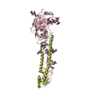
 Components Components | Glycogen phosphorylase, muscle form | ||||||
 Keywords Keywords | TRANSFERASE / glycogenolysis / type 2 diabetes | ||||||
| Function / homology |  Function and homology information Function and homology informationglycogen phosphorylase / glycogen phosphorylase activity / glycogen catabolic process / skeletal muscle myofibril / pyridoxal phosphate binding / nucleotide binding Similarity search - Function | ||||||
| Biological species |  | ||||||
| Method |  X-RAY DIFFRACTION / SYNCHROTRON / FOURIER SYNTHESIS / Resolution: 2.03 Å X-RAY DIFFRACTION / SYNCHROTRON / FOURIER SYNTHESIS / Resolution: 2.03 Å | ||||||
 Authors Authors | Chrysina, E.D. / Kosmopoulou, M.N. / Leonidas, D.D. / Oikonomakos, N.G. | ||||||
 Citation Citation | Journal: Eur.J.Org.Chem. / Year: 2007 Title: In the Search of Glycogen Phosphorylase Inhibitors: Synthesis of C-D-Glycopyranosylbenzo(hydro)quinones Inhibition of and Binding to Glycogen Phosphorylase in the Crystal Authors: He, L. / Zhang, Y.Z. / Tanoh, M. / Chen, G.-R. / Praly, J.-P. / Chrysina, E.D. / Tiraidis, C. / Kosmopoulou, M. / Leonidas, D.D. / Oikonomakos, N.G. | ||||||
| History |
|
- Structure visualization
| Structure viewer | Molecule: MolmilJmol/JSmol |
|---|
- Downloads & links
-Download
| PDBx/mmCIF format | 2fet.cif.gz | 179.4 KB | Display | PDBx/mmCIF format |
|---|---|---|---|---|
| PDB format | pdb2fet.ent.gz | 140.5 KB | Display | PDB format |
| PDBx/mmJSON format | 2fet.json.gz | Tree view | PDBx/mmJSON format | |
| Others |  Other downloads Other downloads |
-Validation report
| Arichive directory | https://data.pdbj.org/pub/pdb/validation_reports/fe/2fetftp://data.pdbj.org/pub/pdb/validation_reports/fe/2fet | HTTPS FTP |
|---|
-Related structure data
| Related structure data | 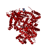 2prjS S: Starting model for refinement |
|---|---|
| Similar structure data |


-Links
 PDBj
PDBj
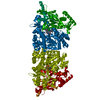
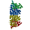
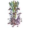
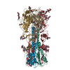
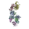
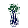
- Assembly
| Deposited unit | 
| ||||||||
|---|---|---|---|---|---|---|---|---|---|
| 1 | 
| ||||||||
| 2 |
| ||||||||
| 3 | 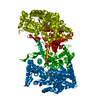
| ||||||||
| Unit cell |
| ||||||||
| Details | Dimeric glycogen phosphorylase is the physiologiacally active species |
-Components
| #1: Protein | Mass: 97291.203 Da / Num. of mol.: 1 / Source method: isolated from a natural source / Details: PYRIDOXAL-5'-PHOSPHATE bound / Source: (natural) |
|---|---|
| #2: Chemical | ChemComp-PLP /   Mass: 247.142 Da / Num. of mol.: 1 / Source method: obtained synthetically / Formula: C8H10NO6P Mass: 247.142 Da / Num. of mol.: 1 / Source method: obtained synthetically / Formula: C8H10NO6P |
| #3: Sugar | ChemComp-H53 / (  Type: D-saccharide / Mass: 272.251 Da / Num. of mol.: 1 / Source method: obtained synthetically / Formula: C12H16O7 Type: D-saccharide / Mass: 272.251 Da / Num. of mol.: 1 / Source method: obtained synthetically / Formula: C12H16O7 |
| #4: Water | ChemComp-HOH /  Mass: 18.015 Da / Num. of mol.: 270 / Source method: isolated from a natural source / Formula: H2O Mass: 18.015 Da / Num. of mol.: 270 / Source method: isolated from a natural source / Formula: H2O |
-Experimental details
-Experiment
| Experiment | Method: X-RAY DIFFRACTION / Number of used crystals: 1 |
|---|
- Sample preparation
| Crystal | Density Matthews: 2.45 Å3/Da / Density % sol: 49.87 % |
|---|---|
| Crystal grow | Temperature: 289 K / Method: small tubes / pH: 6.7 Details: 10 mM Bes buffer, 3 mM DDT, pH 6.7, SMALL TUBES, temperature 289K |
-Data collection
| Diffraction | Mean temperature: 293 K |
|---|---|
| Diffraction source | Source: SYNCHROTRON / Site: EMBL/DESY, HAMBURG  / Beamline: X13 / Wavelength: 0.803 Å / Beamline: X13 / Wavelength: 0.803 Å |
| Detector | Type: ADSC QUANTUM 4 / Detector: CCD / Date: Oct 31, 2003 |
| Radiation | Monochromator: CRYSTAL / Protocol: SINGLE WAVELENGTH / Monochromatic (M) / Laue (L): M / Scattering type: x-ray |
| Radiation wavelength | Wavelength: 0.803 Å / Relative weight: 1 |
| Reflection | Resolution: 2.03→30 Å / Num. all: 56087 / Num. obs: 56087 / % possible obs: 91.7 % / Observed criterion σ(I): -3 / Redundancy: 3.4 % / Biso Wilson estimate: 31.7 Å2 / Rsym value: 0.056 / Net I/σ(I): 13.1 |
| Reflection shell | Resolution: 2.03→2.09 Å / Redundancy: 3.6 % / Mean I/σ(I) obs: 3.6 / Num. unique all: 2654 / Rsym value: 0.486 / % possible all: 88.4 |
- Processing
| Software |
| |||||||||||||||||||||||||
|---|---|---|---|---|---|---|---|---|---|---|---|---|---|---|---|---|---|---|---|---|---|---|---|---|---|---|
| Refinement | Method to determine structure: FOURIER SYNTHESIS Starting model: 2PRJ Resolution: 2.03→30 Å / Isotropic thermal model: RESTRAINED / Cross valid method: THROUGHOUT / σ(F): 0 / Stereochemistry target values: Engh & Huber
| |||||||||||||||||||||||||
| Displacement parameters | Biso mean: 38.7 Å2
| |||||||||||||||||||||||||
| Refine analyze |
| |||||||||||||||||||||||||
| Refinement step | Cycle: LAST / Resolution: 2.03→30 Å
| |||||||||||||||||||||||||
| Refine LS restraints |
| |||||||||||||||||||||||||
| LS refinement shell | Resolution: 2.03→2.1 Å
|
