 Movie
Movie Controller

[English] 日本語
 Yorodumi
- PDB-1yga: CRYSTAL STRUCTURE OF Saccharomyces cerevisiae YN9A PROTEIN, NEW Y... -
+ Open data

- Basic information
| Entry | Database: PDB / ID: 1yga | ||||||
|---|---|---|---|---|---|---|---|
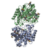
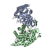
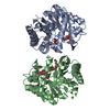
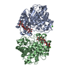
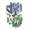
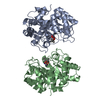
| Title | CRYSTAL STRUCTURE OF Saccharomyces cerevisiae YN9A PROTEIN, NEW YORK STRUCTURAL GENOMICS CONSORTIUM | ||||||
 Components Components | Hypothetical 37.9 kDa protein in BIO3-HXT17 intergenic region | ||||||
 Keywords Keywords | ISOMERASE / ALDOSE_1_EPIMERASE / SUGAR METABOLISM / PREDICTED / STRUCTURAL GENOMICS / PSI / Protein Structure Initiative / New York SGX Research Center for Structural Genomics / NYSGXRC | ||||||
| Function / homology |  Function and homology information Function and homology informationIsomerases / aldose 1-epimerase activity / galactose catabolic process via UDP-galactose, Leloir pathway / glucose metabolic process / carbohydrate binding Similarity search - Function | ||||||
| Biological species |  | ||||||
| Method |  X-RAY DIFFRACTION / MIR / Resolution: 2 Å X-RAY DIFFRACTION / MIR / Resolution: 2 Å | ||||||
 Authors Authors | Patskovsky, Y. / Almo, S.C. / Burley, S.K. / New York SGX Research Center for Structural Genomics (NYSGXRC) | ||||||
 Citation Citation | Journal: To be Published Title: Crystal Structure of the Yeast Aldose_1_Epimerase Authors: Patskovsky, Y. / Almo, S.C. | ||||||
| History |
|
- Structure visualization
| Structure viewer | Molecule: MolmilJmol/JSmol |
|---|
- Downloads & links
-Download
| PDBx/mmCIF format | 1yga.cif.gz | 150.9 KB | Display | PDBx/mmCIF format |
|---|---|---|---|---|
| PDB format | pdb1yga.ent.gz | 118.4 KB | Display | PDB format |
| PDBx/mmJSON format | 1yga.json.gz | Tree view | PDBx/mmJSON format | |
| Others |  Other downloads Other downloads |
-Validation report
| Summary document | 1yga_validation.pdf.gz | 426.4 KB | Display | wwPDB validaton report |
|---|---|---|---|---|
| Full document | 1yga_full_validation.pdf.gz | 433.9 KB | Display | |
| Data in XML | 1yga_validation.xml.gz | 31.1 KB | Display | |
| Data in CIF | 1yga_validation.cif.gz | 46.1 KB | Display | |
| Arichive directory | https://data.pdbj.org/pub/pdb/validation_reports/yg/1ygaftp://data.pdbj.org/pub/pdb/validation_reports/yg/1yga | HTTPS FTP |
-Related structure data
| Similar structure data | |
|---|---|
| Other databases |


-Links
 PDBj
PDBj


- Assembly
| Deposited unit | 
| ||||||||
|---|---|---|---|---|---|---|---|---|---|
| 1 |
| ||||||||
| Unit cell |
| ||||||||
| Details | The biological assembly is a homodimer composed of two identical monomers. The dimeric structure is stabilized by intra-chain S-S bridges. The asymmetric unit contains dimer. |
-Components
| #1: Protein | Mass: 37896.598 Da / Num. of mol.: 2 Source method: isolated from a genetically manipulated source Source: (gene. exp.) Gene: YN9A / Species (production host): Escherichia coli / Production host:  #2: Water | ChemComp-HOH / |  Mass: 18.015 Da / Num. of mol.: 539 / Source method: isolated from a natural source / Formula: H2O Mass: 18.015 Da / Num. of mol.: 539 / Source method: isolated from a natural source / Formula: H2OHas protein modification | Y | |
|---|
-Experimental details
-Experiment
| Experiment | Method: X-RAY DIFFRACTION / Number of used crystals: 2 |
|---|
- Sample preparation
| Crystal | Density Matthews: 2.38 Å3/Da / Density % sol: 49.1 % |
|---|---|
| Crystal grow | Temperature: 290 K / Method: vapor diffusion, hanging drop / pH: 8.5 Details: PEG4000, TRIS-HCL, pH 8.50, VAPOR DIFFUSION, HANGING DROP, temperature 290K |
-Data collection
| Diffraction | Mean temperature: 88 K |
|---|---|
| Diffraction source | Source: ROTATING ANODE / Type: RIGAKU RU200 / Wavelength: 1.5418 / Wavelength: 1.5418 Å |
| Detector | Type: RIGAKU RAXIS IV / Detector: IMAGE PLATE / Date: Dec 20, 2004 / Details: MIRRORS |
| Radiation | Monochromator: MIRRORS / Protocol: SINGLE WAVELENGTH / Monochromatic (M) / Laue (L): M / Scattering type: x-ray |
| Radiation wavelength | Wavelength: 1.5418 Å / Relative weight: 1 |
| Reflection | Resolution: 2→20 Å / Num. all: 44056 / Num. obs: 44056 / % possible obs: 89.4 % / Observed criterion σ(F): 0 / Observed criterion σ(I): 0 / Redundancy: 12.6 % / Biso Wilson estimate: 11.3 Å2 / Rmerge(I) obs: 0.065 / Rsym value: 0.077 / Net I/σ(I): 12.1 |
| Reflection shell | Resolution: 2→2.07 Å / Redundancy: 1.8 % / Rmerge(I) obs: 0.24 / Mean I/σ(I) obs: 2.7 / Num. unique all: 2328 / Rsym value: 0.25 / % possible all: 48.1 |
- Processing
| Software |
| ||||||||||||||||||||||||||||||||||||
|---|---|---|---|---|---|---|---|---|---|---|---|---|---|---|---|---|---|---|---|---|---|---|---|---|---|---|---|---|---|---|---|---|---|---|---|---|---|
| Refinement | Method to determine structure: MIR / Resolution: 2→20 Å / Rfactor Rfree error: 0.006 / Data cutoff high absF: 242583.3 / Data cutoff low absF: 0 / Isotropic thermal model: RESTRAINED / Cross valid method: THROUGHOUT / σ(F): 1 / Stereochemistry target values: Engh & Huber
| ||||||||||||||||||||||||||||||||||||
| Solvent computation | Solvent model: FLAT MODEL / Bsol: 27.0966 Å2 / ksol: 0.324547 e/Å3 | ||||||||||||||||||||||||||||||||||||
| Displacement parameters | Biso mean: 25.6 Å2
| ||||||||||||||||||||||||||||||||||||
| Refine analyze |
| ||||||||||||||||||||||||||||||||||||
| Refinement step | Cycle: LAST / Resolution: 2→20 Å
| ||||||||||||||||||||||||||||||||||||
| Refine LS restraints |
| ||||||||||||||||||||||||||||||||||||
| LS refinement shell | Resolution: 2→2.13 Å / Rfactor Rfree error: 0.025 / Total num. of bins used: 6
| ||||||||||||||||||||||||||||||||||||
| Xplor file |
|
