 Movie
Movie Controller

[English] 日本語
 Yorodumi
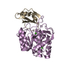
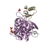
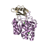
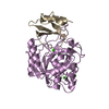
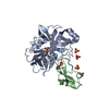
- PDB-1y4d: Crystal structure of the complex of subtilisin BPN' with chymotry... -
+ Open data

- Basic information
| Entry | Database: PDB / ID: 1y4d | ||||||
|---|---|---|---|---|---|---|---|
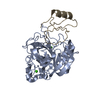
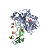
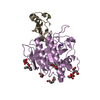
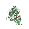
| Title | Crystal structure of the complex of subtilisin BPN' with chymotrypsin inhibitor 2 M59R/E60S mutant | ||||||
 Components Components |
| ||||||
 Keywords Keywords | HYDROLASE/HYDROLASE INHIBITOR / serine protease / inhibitor / HYDROLASE-HYDROLASE INHIBITOR COMPLEX | ||||||
| Function / homology |  Function and homology information Function and homology informationsubtilisin / sporulation resulting in formation of a cellular spore / fibrinolysis / serine-type endopeptidase inhibitor activity / response to wounding / serine-type endopeptidase activity / proteolysis / extracellular region / metal ion binding Similarity search - Function | ||||||
| Biological species |   | ||||||
| Method |  X-RAY DIFFRACTION / SYNCHROTRON / MOLECULAR REPLACEMENT / Resolution: 2 Å X-RAY DIFFRACTION / SYNCHROTRON / MOLECULAR REPLACEMENT / Resolution: 2 Å | ||||||
 Authors Authors | Radisky, E.S. / Lu, C.J. / Kwan, G. / Koshland Jr., D.E. | ||||||
 Citation Citation | Journal: Biochemistry / Year: 2005 Title: Role of the intramolecular hydrogen bond network in the inhibitory power of chymotrypsin inhibitor 2 Authors: Radisky, E.S. / Lu, C.J. / Kwan, G. / Koshland Jr., D.E. | ||||||
| History |
|
- Structure visualization
| Structure viewer | Molecule: MolmilJmol/JSmol |
|---|
- Downloads & links
-Download
| PDBx/mmCIF format | 1y4d.cif.gz | 79 KB | Display | PDBx/mmCIF format |
|---|---|---|---|---|
| PDB format | pdb1y4d.ent.gz | 57.9 KB | Display | PDB format |
| PDBx/mmJSON format | 1y4d.json.gz | Tree view | PDBx/mmJSON format | |
| Others |  Other downloads Other downloads |
-Validation report
| Summary document | 1y4d_validation.pdf.gz | 437.4 KB | Display | wwPDB validaton report |
|---|---|---|---|---|
| Full document | 1y4d_full_validation.pdf.gz | 441.4 KB | Display | |
| Data in XML | 1y4d_validation.xml.gz | 15.4 KB | Display | |
| Data in CIF | 1y4d_validation.cif.gz | 21.8 KB | Display | |
| Arichive directory | https://data.pdbj.org/pub/pdb/validation_reports/y4/1y4dftp://data.pdbj.org/pub/pdb/validation_reports/y4/1y4d | HTTPS FTP |
-Related structure data
| Related structure data | 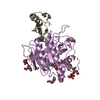 1y1kC 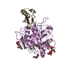 1y33C 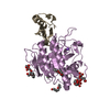 1y34C 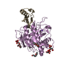 1y3bC 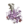 1y3cC 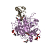 1y3dC 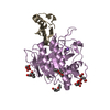 1y3fC  1y48C 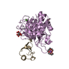 1y4aC  1tm3S C: citing same article ( S: Starting model for refinement |
|---|---|
| Similar structure data |

-Links
 PDBj
PDBj

- Assembly
| Deposited unit | 
| ||||||||||
|---|---|---|---|---|---|---|---|---|---|---|---|
| 1 |
| ||||||||||
| Unit cell |
|
-Components
| #1: Protein | Mass: 28381.396 Da / Num. of mol.: 1 / Mutation: C-terminal 6-His tag Source method: isolated from a genetically manipulated source Source: (gene. exp.) |
|---|---|
| #2: Protein | Mass: 7267.563 Da / Num. of mol.: 1 / Mutation: initiating Met, M59R/E60S Source method: isolated from a genetically manipulated source Source: (gene. exp.) |
| #3: Chemical | ChemComp-CA /   Mass: 40.078 Da / Num. of mol.: 1 / Source method: obtained synthetically / Formula: Ca Mass: 40.078 Da / Num. of mol.: 1 / Source method: obtained synthetically / Formula: Ca |
| #4: Chemical | ChemComp-NA /   Mass: 22.990 Da / Num. of mol.: 1 / Source method: obtained synthetically / Formula: Na Mass: 22.990 Da / Num. of mol.: 1 / Source method: obtained synthetically / Formula: Na |
| #5: Water | ChemComp-HOH /  Mass: 18.015 Da / Num. of mol.: 114 / Source method: isolated from a natural source / Formula: H2O Mass: 18.015 Da / Num. of mol.: 114 / Source method: isolated from a natural source / Formula: H2O |
-Experimental details
-Experiment
| Experiment | Method: X-RAY DIFFRACTION / Number of used crystals: 1 |
|---|
- Sample preparation
| Crystal | Density Matthews: 2.3 Å3/Da / Density % sol: 45.7 % |
|---|---|
| Crystal grow | Temperature: 277 K / Method: vapor diffusion, hanging drop / pH: 4.5 Details: potassium phosphate, PEG 8000, pH 4.5, VAPOR DIFFUSION, HANGING DROP, temperature 277K |
-Data collection
| Diffraction | Mean temperature: 100 K |
|---|---|
| Diffraction source | Source: SYNCHROTRON / Site: ALS  / Beamline: 8.3.1 / Wavelength: 1 Å / Beamline: 8.3.1 / Wavelength: 1 Å |
| Detector | Type: ADSC QUANTUM 4 / Detector: CCD / Date: Jun 4, 2003 |
| Radiation | Protocol: SINGLE WAVELENGTH / Monochromatic (M) / Laue (L): M / Scattering type: x-ray |
| Radiation wavelength | Wavelength: 1 Å / Relative weight: 1 |
| Reflection | Resolution: 2→32.55 Å / Num. all: 22160 / Num. obs: 22160 / % possible obs: 99.6 % / Observed criterion σ(I): -3 / Redundancy: 4.9 % / Rsym value: 0.157 / Net I/σ(I): 7.4 |
- Processing
| Software |
| |||||||||||||||||||||||||||||||||||||||||||||||||||||||||||||||||||||||||||
|---|---|---|---|---|---|---|---|---|---|---|---|---|---|---|---|---|---|---|---|---|---|---|---|---|---|---|---|---|---|---|---|---|---|---|---|---|---|---|---|---|---|---|---|---|---|---|---|---|---|---|---|---|---|---|---|---|---|---|---|---|---|---|---|---|---|---|---|---|---|---|---|---|---|---|---|---|
| Refinement | Method to determine structure: MOLECULAR REPLACEMENT Starting model: 1TM3 Resolution: 2→32.55 Å / Cor.coef. Fo:Fc: 0.94 / Cor.coef. Fo:Fc free: 0.906 / SU B: 4.621 / SU ML: 0.129 / Cross valid method: THROUGHOUT / σ(F): 0 / ESU R: 0.197 / ESU R Free: 0.18 / Stereochemistry target values: MAXIMUM LIKELIHOOD
| |||||||||||||||||||||||||||||||||||||||||||||||||||||||||||||||||||||||||||
| Solvent computation | Ion probe radii: 0.8 Å / Shrinkage radii: 0.8 Å / VDW probe radii: 1.4 Å / Solvent model: BABINET MODEL WITH MASK | |||||||||||||||||||||||||||||||||||||||||||||||||||||||||||||||||||||||||||
| Displacement parameters | Biso mean: 26.581 Å2
| |||||||||||||||||||||||||||||||||||||||||||||||||||||||||||||||||||||||||||
| Refinement step | Cycle: LAST / Resolution: 2→32.55 Å
| |||||||||||||||||||||||||||||||||||||||||||||||||||||||||||||||||||||||||||
| Refine LS restraints |
| |||||||||||||||||||||||||||||||||||||||||||||||||||||||||||||||||||||||||||
| LS refinement shell | Resolution: 2→2.052 Å / Total num. of bins used: 20 /
|
