 Movie
Movie Controller

[English] 日本語
 Yorodumi
- PDB-1xuv: X-Ray Crystal Structure of Protein MM0500 from Methanosarcina maz... -
+ Open data

- Basic information
| Entry | Database: PDB / ID: 1xuv | ||||||
|---|---|---|---|---|---|---|---|
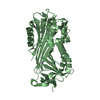
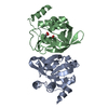
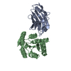
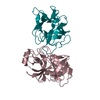
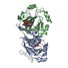
| Title | X-Ray Crystal Structure of Protein MM0500 from Methanosarcina mazei. Northeast Structural Genomics Consortium Target MaR10. | ||||||
 Components Components | hypothetical protein MM0500 | ||||||
 Keywords Keywords | STRUCTURAL GENOMICS / UNKNOWN FUNCTION / Alpha-beta protein / Northeast Structural Genomics Consortium / NESG / Protein Structure Initiative / PSI | ||||||
| Function / homology | Activator of Hsp90 ATPase homologue 1-like / Activator of Hsp90 ATPase homolog 1-like protein / START domain / Alpha-D-Glucose-1,6-Bisphosphate; Chain A, domain 4 / START-like domain superfamily / 2-Layer Sandwich / Alpha Beta / Activator of Hsp90 ATPase homologue 1/2-like C-terminal domain-containing protein Function and homology information Function and homology information | ||||||
| Biological species |  Methanosarcina mazei Go1 (archaea) Methanosarcina mazei Go1 (archaea) | ||||||
| Method |  X-RAY DIFFRACTION / SYNCHROTRON / SAD / Resolution: 2.1 Å X-RAY DIFFRACTION / SYNCHROTRON / SAD / Resolution: 2.1 Å | ||||||
 Authors Authors | Forouhar, F. / Abashidze, M. / Ciano, M. / Acton, T.B. / Montelione, G.T. / Tong, L. / Hunt, J.F. / Northeast Structural Genomics Consortium (NESG) | ||||||
 Citation Citation | Journal: To be Published Title: Crystal Structure of the Hypothetical Protein from Methanosarcina mazei, Northeast Strcutural Genomics Target MaR10 Authors: Forouhar, F. / Abashidze, M. / Ciano, M. / Acton, T.B. / Montelione, G.T. / Tong, L. / Hunt, J.F. | ||||||
| History |
|
- Structure visualization
| Structure viewer | Molecule: MolmilJmol/JSmol |
|---|
- Downloads & links
-Download
| PDBx/mmCIF format | 1xuv.cif.gz | 114.9 KB | Display | PDBx/mmCIF format |
|---|---|---|---|---|
| PDB format | pdb1xuv.ent.gz | 90.1 KB | Display | PDB format |
| PDBx/mmJSON format | 1xuv.json.gz | Tree view | PDBx/mmJSON format | |
| Others |  Other downloads Other downloads |
-Validation report
| Summary document | 1xuv_validation.pdf.gz | 448.1 KB | Display | wwPDB validaton report |
|---|---|---|---|---|
| Full document | 1xuv_full_validation.pdf.gz | 460 KB | Display | |
| Data in XML | 1xuv_validation.xml.gz | 23.5 KB | Display | |
| Data in CIF | 1xuv_validation.cif.gz | 32.1 KB | Display | |
| Arichive directory | https://data.pdbj.org/pub/pdb/validation_reports/xu/1xuvftp://data.pdbj.org/pub/pdb/validation_reports/xu/1xuv | HTTPS FTP |
-Related structure data
| Similar structure data | |
|---|---|
| Other databases |

-Links
 PDBj
PDBj- Assembly
| Deposited unit | 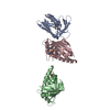
| ||||||||
|---|---|---|---|---|---|---|---|---|---|
| 1 | 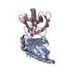
| ||||||||
| 2 | 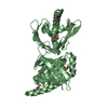
| ||||||||
| Unit cell |
| ||||||||
| Details | possibly dimer |
-Components
| #1: Protein | Mass: 20695.639 Da / Num. of mol.: 3 Source method: isolated from a genetically manipulated source Source: (gene. exp.) Methanosarcina mazei Go1 (archaea) / Species: Methanosarcina mazei / Strain: Goe1 / Plasmid: BL21(DE3)+Magic / Production host:  #2: Water | ChemComp-HOH / |  Mass: 18.015 Da / Num. of mol.: 247 / Source method: isolated from a natural source / Formula: H2O Mass: 18.015 Da / Num. of mol.: 247 / Source method: isolated from a natural source / Formula: H2OHas protein modification | Y | |
|---|
-Experimental details
-Experiment
| Experiment | Method: X-RAY DIFFRACTION / Number of used crystals: 1 |
|---|
- Sample preparation
| Crystal | Density Matthews: 2.5 Å3/Da / Density % sol: 50 % |
|---|---|
| Crystal grow | Temperature: 293 K / Method: vapor diffusion, hanging drop / pH: 4.6 Details: 100 mM Acetate (pH 4.6), 8% PEG 4k, 5 mM DTT, VAPOR DIFFUSION, HANGING DROP, temperature 293K |
-Data collection
| Diffraction | Mean temperature: 100 K |
|---|---|
| Diffraction source | Source: SYNCHROTRON / Site: NSLS  / Beamline: X4A / Wavelength: 0.97916 Å / Beamline: X4A / Wavelength: 0.97916 Å |
| Detector | Type: ADSC QUANTUM 4 / Detector: CCD / Date: Oct 14, 2004 / Details: mirrors |
| Radiation | Monochromator: Si 111 CHANNEL / Protocol: SINGLE WAVELENGTH / Monochromatic (M) / Laue (L): M / Scattering type: x-ray |
| Radiation wavelength | Wavelength: 0.97916 Å / Relative weight: 1 |
| Reflection | Resolution: 2.1→30 Å / Num. all: 67882 / Num. obs: 66458 / % possible obs: 99 % / Observed criterion σ(F): 2 / Observed criterion σ(I): 2 / Redundancy: 4.4 % / Biso Wilson estimate: 13.1 Å2 / Rmerge(I) obs: 0.042 / Rsym value: 0.035 / Net I/σ(I): 29.6 |
| Reflection shell | Resolution: 2.1→2.18 Å / Redundancy: 4.4 % / Rmerge(I) obs: 0.27 / Mean I/σ(I) obs: 5.6 / Num. unique all: 6697 / Rsym value: 0.211 / % possible all: 99.9 |
- Processing
| Software |
| |||||||||||||||||||||||||
|---|---|---|---|---|---|---|---|---|---|---|---|---|---|---|---|---|---|---|---|---|---|---|---|---|---|---|
| Refinement | Method to determine structure: SAD / Resolution: 2.1→28.59 Å / Rfactor Rfree error: 0.003 / Data cutoff high absF: 489385.78 / Data cutoff low absF: 0 / Isotropic thermal model: OVERALL / Cross valid method: THROUGHOUT / σ(F): 2 / σ(I): 2 / Stereochemistry target values: Engh & Huber
| |||||||||||||||||||||||||
| Solvent computation | Solvent model: FLAT MODEL / Bsol: 46.9852 Å2 / ksol: 0.338881 e/Å3 | |||||||||||||||||||||||||
| Displacement parameters | Biso mean: 43.1 Å2
| |||||||||||||||||||||||||
| Refine analyze |
| |||||||||||||||||||||||||
| Refinement step | Cycle: LAST / Resolution: 2.1→28.59 Å
| |||||||||||||||||||||||||
| Refine LS restraints |
| |||||||||||||||||||||||||
| LS refinement shell | Resolution: 2.1→2.23 Å / Rfactor Rfree error: 0.01 / Total num. of bins used: 6
|
