 Movie
Movie Controller


+ Open data

- Basic information
| Entry | Database: PDB / ID: 3cry | ||||||
|---|---|---|---|---|---|---|---|
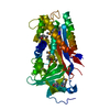
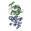
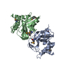
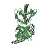
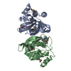
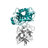
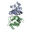
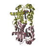
| Title | Gamma-glutamyl cyclotransferase | ||||||
 Components Components | Gamma-glutamyl cyclotransferase | ||||||
 Keywords Keywords | TRANSFERASE / enzyme / cyclotransferase / gamma-glutamyl / oxoproline | ||||||
| Function / homology |  Function and homology information Function and homology informationgamma-glutamylcyclotransferase / gamma-glutamylcyclotransferase activity / Glutathione synthesis and recycling / release of cytochrome c from mitochondria / protein homodimerization activity / extracellular exosome / cytosol Similarity search - Function | ||||||
| Biological species |  Homo sapiens (human) Homo sapiens (human) | ||||||
| Method |  X-RAY DIFFRACTION / SYNCHROTRON / MOLECULAR REPLACEMENT / Resolution: 1.7 Å X-RAY DIFFRACTION / SYNCHROTRON / MOLECULAR REPLACEMENT / Resolution: 1.7 Å | ||||||
 Authors Authors | Oakley, A.J. / Board, P.G. | ||||||
 Citation Citation | Journal: J.Biol.Chem. / Year: 2008 Title: The identification and structural characterization of C7orf24 as gamma-glutamyl cyclotransferase. An essential enzyme in the gamma-glutamyl cycle Authors: Oakley, A.J. / Yamada, T. / Liu, D. / Coggan, M. / Clark, A.G. / Board, P.G. | ||||||
| History |
|
- Structure visualization
| Structure viewer | Molecule: MolmilJmol/JSmol |
|---|
- Downloads & links
-Download
| PDBx/mmCIF format | 3cry.cif.gz | 92.6 KB | Display | PDBx/mmCIF format |
|---|---|---|---|---|
| PDB format | pdb3cry.ent.gz | 70.1 KB | Display | PDB format |
| PDBx/mmJSON format | 3cry.json.gz | Tree view | PDBx/mmJSON format | |
| Others |  Other downloads Other downloads |
-Validation report
| Arichive directory | https://data.pdbj.org/pub/pdb/validation_reports/cr/3cryftp://data.pdbj.org/pub/pdb/validation_reports/cr/3cry | HTTPS FTP |
|---|
-Related structure data
| Related structure data |  2pn7C  2rbhSC C: citing same article ( S: Starting model for refinement |
|---|---|
| Similar structure data |

-Links
 PDBj
PDBj- Assembly
| Deposited unit | 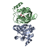
| ||||||||
|---|---|---|---|---|---|---|---|---|---|
| 1 |
| ||||||||
| Unit cell |
|
-Components
| #1: Protein | Mass: 21028.705 Da / Num. of mol.: 2 / Mutation: E98Q Source method: isolated from a genetically manipulated source Source: (gene. exp.) Homo sapiens (human) / Gene: GGCT / Plasmid: pHUE / Species (production host): Escherichia coli / Production host:  #2: Chemical |   Mass: 59.044 Da / Num. of mol.: 3 / Source method: obtained synthetically / Formula: C2H3O2 Mass: 59.044 Da / Num. of mol.: 3 / Source method: obtained synthetically / Formula: C2H3O2#3: Water | ChemComp-HOH / |  Mass: 18.015 Da / Num. of mol.: 399 / Source method: isolated from a natural source / Formula: H2O Mass: 18.015 Da / Num. of mol.: 399 / Source method: isolated from a natural source / Formula: H2O |
|---|
-Experimental details
-Experiment
| Experiment | Method: X-RAY DIFFRACTION |
|---|
- Sample preparation
| Crystal | Density Matthews: 2.04 Å3/Da / Density % sol: 39.76 % |
|---|---|
| Crystal grow | Temperature: 277 K / Method: vapor diffusion, hanging drop / pH: 5 Details: 50% PEG 4000, 0.2M ammonium acetate, 0.1M sodium acetate, pH 5.0, VAPOR DIFFUSION, HANGING DROP, temperature 277K |
-Data collection
| Diffraction | Mean temperature: 100 K |
|---|---|
| Diffraction source | Source: SYNCHROTRON / Site: Australian Synchrotron  / Beamline: MX1 / Wavelength: 0.95361 Å / Beamline: MX1 / Wavelength: 0.95361 Å |
| Detector | Type: MAR CCD 165 mm / Detector: CCD / Date: Nov 15, 2007 / Details: mirrors |
| Radiation | Monochromator: BEAMLINE OPTICS / Protocol: SINGLE WAVELENGTH / Monochromatic (M) / Laue (L): M / Scattering type: x-ray |
| Radiation wavelength | Wavelength: 0.95361 Å / Relative weight: 1 |
| Reflection | Resolution: 1.7→70.19 Å / Num. all: 38869 / Num. obs: 38628 / % possible obs: 99.4 % / Observed criterion σ(F): -2 / Observed criterion σ(I): -2 / Redundancy: 4.6 % / Biso Wilson estimate: 13.73 Å2 / Rmerge(I) obs: 0.092 / Net I/σ(I): 12.6 |
| Reflection shell | Resolution: 1.7→1.8 Å / Redundancy: 4.4 % / Rmerge(I) obs: 0.419 / Mean I/σ(I) obs: 3.7 / Num. unique all: 5449 / % possible all: 97.7 |
- Processing
| Software |
| ||||||||||||||||||||||||||||||||||||||||||||||||||||||||||||||||||||||||||||||||||||||||||
|---|---|---|---|---|---|---|---|---|---|---|---|---|---|---|---|---|---|---|---|---|---|---|---|---|---|---|---|---|---|---|---|---|---|---|---|---|---|---|---|---|---|---|---|---|---|---|---|---|---|---|---|---|---|---|---|---|---|---|---|---|---|---|---|---|---|---|---|---|---|---|---|---|---|---|---|---|---|---|---|---|---|---|---|---|---|---|---|---|---|---|---|
| Refinement | Method to determine structure: MOLECULAR REPLACEMENT Starting model: 2RBH Resolution: 1.7→22.37 Å / Cor.coef. Fo:Fc: 0.952 / Cor.coef. Fo:Fc free: 0.926 / SU B: 2.05 / SU ML: 0.07 / Isotropic thermal model: Isotropic / Cross valid method: THROUGHOUT / σ(F): -1 / σ(I): -1 / ESU R: 0.114 / ESU R Free: 0.115 / Stereochemistry target values: MAXIMUM LIKELIHOOD / Details: HYDROGENS HAVE BEEN ADDED IN THE RIDING POSITIONS
| ||||||||||||||||||||||||||||||||||||||||||||||||||||||||||||||||||||||||||||||||||||||||||
| Solvent computation | Ion probe radii: 0.8 Å / Shrinkage radii: 0.8 Å / VDW probe radii: 1.2 Å / Solvent model: MASK | ||||||||||||||||||||||||||||||||||||||||||||||||||||||||||||||||||||||||||||||||||||||||||
| Displacement parameters | Biso mean: 14.957 Å2
| ||||||||||||||||||||||||||||||||||||||||||||||||||||||||||||||||||||||||||||||||||||||||||
| Refinement step | Cycle: LAST / Resolution: 1.7→22.37 Å
| ||||||||||||||||||||||||||||||||||||||||||||||||||||||||||||||||||||||||||||||||||||||||||
| Refine LS restraints |
| ||||||||||||||||||||||||||||||||||||||||||||||||||||||||||||||||||||||||||||||||||||||||||
| LS refinement shell | Resolution: 1.7→1.744 Å / Total num. of bins used: 20
|
