[English] 日本語
 Yorodumi
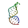
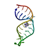
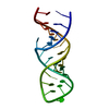
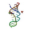
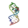
- PDB-1xsu: Solution structure of E.coli RNase P RNA P4 stem, U69C/C70U mutat... -
+ Open data
- Basic information
| Entry | Database: PDB / ID: 1xsu | ||||||
|---|---|---|---|---|---|---|---|
| Title | Solution structure of E.coli RNase P RNA P4 stem, U69C/C70U mutation, complexed with cobalt (III) hexammine. | ||||||
 Components Components | RNA (27-MER) | ||||||
 Keywords Keywords | RNA / Ribonuclease P RNA / Ribozyme / transfer RNA processing / P4 stem / U69C/C70U mutant / metal binding site / metal complex / cobalt (III) hexammine complex | ||||||
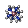
| Function / homology | COBALT HEXAMMINE(III) / RNA / RNA (> 10) Function and homology information Function and homology information | ||||||
| Method | SOLUTION NMR / restrained molecular dynamics, simulated annealing | ||||||
| Model type details | minimized average | ||||||
 Authors Authors | Schmitz, M. | ||||||
 Citation Citation |  Journal: Nucleic Acids Res. / Year: 2004 Journal: Nucleic Acids Res. / Year: 2004Title: Change of RNase P RNA function by single base mutation correlates with perturbation of metal ion binding in P4 as determined by NMR spectroscopy Authors: Schmitz, M. | ||||||
| History |
|
- Structure visualization
| Structure viewer | Molecule:  Molmil Molmil Jmol/JSmol Jmol/JSmol |
|---|
- Downloads & links
- Download
| PDBx/mmCIF format |  1xsu.cif.gz 1xsu.cif.gz | 26.7 KB | Display |  PDBx/mmCIF format PDBx/mmCIF format |
|---|---|---|---|---|
| PDB format |  pdb1xsu.ent.gz pdb1xsu.ent.gz | 17.5 KB | Display |  PDB format PDB format |
| PDBx/mmJSON format |  1xsu.json.gz 1xsu.json.gz | Tree view |  PDBx/mmJSON format PDBx/mmJSON format | |
| Others |  Other downloads Other downloads |
-Validation report
| Arichive directory |  https://data.pdbj.org/pub/pdb/validation_reports/xs/1xsu https://data.pdbj.org/pub/pdb/validation_reports/xs/1xsu ftp://data.pdbj.org/pub/pdb/validation_reports/xs/1xsu ftp://data.pdbj.org/pub/pdb/validation_reports/xs/1xsu | HTTPS FTP |
|---|
-Related structure data
- Links
Links
- Assembly
| Deposited unit | 
| |||||||||
|---|---|---|---|---|---|---|---|---|---|---|
| 1 |
| |||||||||
| NMR ensembles |
|
- Components
| #1: RNA chain | Mass: 8633.143 Da / Num. of mol.: 1 / Mutation: U69C, C70U / Source method: obtained synthetically Details: enzymatically synthesized from DNA oligonucleotide template ny T7 RNA polymerase |
|---|---|
| #2: Chemical | ChemComp-NCO / |
-Experimental details
-Experiment
| Experiment | Method: SOLUTION NMR | ||||||||||||||||||||||||||||
|---|---|---|---|---|---|---|---|---|---|---|---|---|---|---|---|---|---|---|---|---|---|---|---|---|---|---|---|---|---|
| NMR experiment |
| ||||||||||||||||||||||||||||
| NMR details | Text: The structure was determined using standard 2D homonuclear techniques as well as 13C and 31P heteronuclear experiments performed at natural abundance. Intermolecular NOE crosspeaks between RNA ...Text: The structure was determined using standard 2D homonuclear techniques as well as 13C and 31P heteronuclear experiments performed at natural abundance. Intermolecular NOE crosspeaks between RNA protons and cobalt (III) hexammine protons and intermolecular distance constraints derived thereof were used to determine the site of cobalt (III) hexammine binding. |
- Sample preparation
| Details |
| |||||||||||||||
|---|---|---|---|---|---|---|---|---|---|---|---|---|---|---|---|---|
| Sample conditions |
|
-NMR measurement
| Radiation | Protocol: SINGLE WAVELENGTH / Monochromatic (M) / Laue (L): M |
|---|---|
| Radiation wavelength | Relative weight: 1 |
| NMR spectrometer | Type: Bruker DMX / Manufacturer: Bruker / Model: DMX / Field strength: 600 MHz |
- Processing
| NMR software |
| ||||||||||||||||||||
|---|---|---|---|---|---|---|---|---|---|---|---|---|---|---|---|---|---|---|---|---|---|
| Refinement | Method: restrained molecular dynamics, simulated annealing / Software ordinal: 1 Details: The average structure is based on the superposition of 18 structures after refinement. The average RMS deviation between the ensemble and the average structure is 1.87 Angstrom. A total of ...Details: The average structure is based on the superposition of 18 structures after refinement. The average RMS deviation between the ensemble and the average structure is 1.87 Angstrom. A total of 261 NOE-derived distance constraints, 246 dihedral constraints and 48 distance constraints from hydrogen bonds were used in refinement. 12 NOE derived intermolecular distance constraints were used to localize the bound cobalt (III) hexammine. | ||||||||||||||||||||
| NMR representative | Selection criteria: minimized average structure | ||||||||||||||||||||
| NMR ensemble | Conformers submitted total number: 1 |
 Movie
Movie Controller










 PDBj
PDBj



 NMRPipe