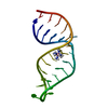
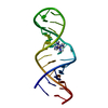
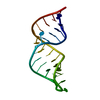
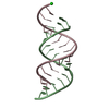
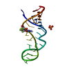
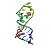
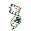
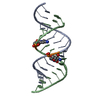
ジャーナル: Nucleic Acids Res. / 年: 2004 タイトル: Change of RNase P RNA function by single base mutation correlates with perturbation of metal ion binding in P4 as determined by NMR spectroscopy 著者: Schmitz, M.
分子量: 8656.183 Da / 分子数: 1 / 由来タイプ: 合成 詳細: enzymatically synthesized from DNA oligonucleotide template by T7 RNA polymerase
-
実験情報
-
実験
実験
手法: 溶液NMR
NMR実験
Conditions-ID
Experiment-ID
Solution-ID
タイプ
1
1
1
2D NOESY
1
2
2
2D NOESY
1
3
2
DQF-COSY
1
4
2
31P-1H-HETERO-COSY
1
5
2
31P-1H-HETERO-TOCSY-NOESY
NMR実験の詳細
Text: The structure was determined using standard 2D homonuclear techniques as well as 13C and 31P heteronuclear experiments performed at natural abundance
タイプ: Bruker DMX / 製造業者: Bruker / モデル: DMX / 磁場強度: 500 MHz
-
解析
NMR software
名称
バージョン
開発者
分類
XwinNMR
1.2
Bruker
collection
NMRPipe
2.1
F. Delaglio
解析
X-PLOR
3.1
構造決定
X-PLOR
3.1
精密化
精密化
手法: restrained molecular dynamics, simulated annealing / ソフトェア番号: 1 詳細: The average structure is based on the superposition of 12 structures after refinement. The average RMS deviation between the ensemble and the average structure is 1.78 Angstrom. A total of ...詳細: The average structure is based on the superposition of 12 structures after refinement. The average RMS deviation between the ensemble and the average structure is 1.78 Angstrom. A total of 290 NOE-derived distance constraints, 245 dihedral restraints and 48 distance restraints from hydrogen bonds were used in refinement.
 ムービー
ムービー コントローラー


 データを開く
 基本情報
基本情報 要素
要素 キーワード
キーワード 機能・相同性情報
 データ登録者
 引用
引用 構造の表示
構造の表示 ダウンロードとリンク
 その他のダウンロード
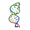


 PDBj
PDBj


 集合体
集合体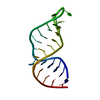
 試料調製
試料調製 解析
解析 NMRPipe