 Movie
Movie Controller

[English] 日本語
 Yorodumi
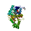
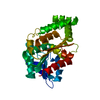
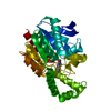
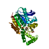
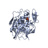
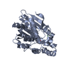
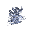
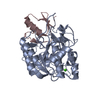
- PDB-1xqy: Crystal structure of F1-mutant S105A complex with PRO-LEU-GLY-GLY -
+ Open data

- Basic information
| Entry | Database: PDB / ID: 1xqy | ||||||
|---|---|---|---|---|---|---|---|
| Title | Crystal structure of F1-mutant S105A complex with PRO-LEU-GLY-GLY | ||||||
 Components Components |
| ||||||
 Keywords Keywords | HYDROLASE / ALPHA-BETA HYDROLASE / CAGED ACTIVE SITE / CATALYTIC TRIAD / NUCLEOPHILE / PEPTIDE CLEAVAGE | ||||||
| Function / homology |  Function and homology information Function and homology informationprolyl aminopeptidase / aminopeptidase activity / proteolysis / membrane Similarity search - Function | ||||||
| Biological species |   Thermoplasma acidophilum (acidophilic) Thermoplasma acidophilum (acidophilic) | ||||||
| Method |  X-RAY DIFFRACTION / MOLECULAR REPLACEMENT / Resolution: 3.2 Å X-RAY DIFFRACTION / MOLECULAR REPLACEMENT / Resolution: 3.2 Å | ||||||
 Authors Authors | Goettig, P. / Brandstetter, H. / Groll, M. / Goehring, W. / Konarev, P.V. / Svergun, D.I. / Huber, R. / Kim, J.-S. | ||||||
 Citation Citation | Journal: J.Biol.Chem. / Year: 2005 Title: X-ray snapshots of peptide processing in mutants of tricorn-interacting factor F1 from Thermoplasma acidophilum Authors: Goettig, P. / Brandstetter, H. / Groll, M. / Goehring, W. / Konarev, P.V. / Svergun, D.I. / Huber, R. / Kim, J.-S. | ||||||
| History |
|
- Structure visualization
| Structure viewer | Molecule: MolmilJmol/JSmol |
|---|
- Downloads & links
-Download
| PDBx/mmCIF format | 1xqy.cif.gz | 72.2 KB | Display | PDBx/mmCIF format |
|---|---|---|---|---|
| PDB format | pdb1xqy.ent.gz | 53.4 KB | Display | PDB format |
| PDBx/mmJSON format | 1xqy.json.gz | Tree view | PDBx/mmJSON format | |
| Others |  Other downloads Other downloads |
-Validation report
| Arichive directory | https://data.pdbj.org/pub/pdb/validation_reports/xq/1xqyftp://data.pdbj.org/pub/pdb/validation_reports/xq/1xqy | HTTPS FTP |
|---|
-Related structure data
| Related structure data |  1xqvC  1xqwC  1xqxC  1xrlC 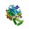 1xrmC 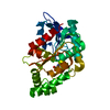 1xrnC 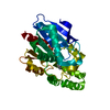 1xroC  1xrpC 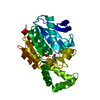 1xrqC  1xrrC C: citing same article ( |
|---|---|
| Similar structure data |


-Links
 PDBj
PDBj


- Assembly
| Deposited unit | 
| ||||||||
|---|---|---|---|---|---|---|---|---|---|
| 1 |
| ||||||||
| Unit cell |
|
-Components
| #1: Protein | Mass: 33514.090 Da / Num. of mol.: 1 / Mutation: S105A Source method: isolated from a genetically manipulated source Source: (gene. exp.) Thermoplasma acidophilum (acidophilic) / Gene: TA0830 / Plasmid: PRSET6C / Production host:  |
|---|---|
| #2: Protein/peptide | Mass: 342.391 Da / Num. of mol.: 1 / Source method: obtained synthetically / Details: chemically synthesized |
| #3: Chemical | ChemComp-PRO /   Type: L-peptide linking / Mass: 115.130 Da / Num. of mol.: 1 / Source method: obtained synthetically / Formula: C5H9NO2 Type: L-peptide linking / Mass: 115.130 Da / Num. of mol.: 1 / Source method: obtained synthetically / Formula: C5H9NO2 |
| #4: Water | ChemComp-HOH /  Mass: 18.015 Da / Num. of mol.: 41 / Source method: isolated from a natural source / Formula: H2O Mass: 18.015 Da / Num. of mol.: 41 / Source method: isolated from a natural source / Formula: H2O |
| Has protein modification | Y |
-Experimental details
-Experiment
| Experiment | Method: X-RAY DIFFRACTION / Number of used crystals: 1 |
|---|
- Sample preparation
| Crystal | Density Matthews: 2.18 Å3/Da / Density % sol: 43.18 % |
|---|---|
| Crystal grow | Temperature: 293 K / Method: vapor diffusion, hanging drop / pH: 6 Details: PEG 6000, Bis-Tris, pH 6.0, VAPOR DIFFUSION, HANGING DROP, temperature 293.0K |
-Data collection
| Diffraction | Mean temperature: 100 K |
|---|---|
| Diffraction source | Source: ROTATING ANODE / Type: RIGAKU RU300 / Wavelength: 1.5418 Å |
| Detector | Type: MARRESEARCH / Detector: IMAGE PLATE / Date: Jul 16, 2003 / Details: MIRRORS |
| Radiation | Protocol: SINGLE WAVELENGTH / Monochromatic (M) / Laue (L): M / Scattering type: x-ray |
| Radiation wavelength | Wavelength: 1.5418 Å / Relative weight: 1 |
| Reflection | Resolution: 2.8→19.7 Å / Num. all: 5742 / Num. obs: 5642 / % possible obs: 98.3 % / Observed criterion σ(F): 1 / Observed criterion σ(I): 1 / Net I/σ(I): 10.1 |
| Reflection shell | Resolution: 2.83→3 Å / % possible all: 55.8 |
- Processing
| Software |
| |||||||||||||||||||||||||
|---|---|---|---|---|---|---|---|---|---|---|---|---|---|---|---|---|---|---|---|---|---|---|---|---|---|---|
| Refinement | Method to determine structure: MOLECULAR REPLACEMENT / Resolution: 3.2→19.66 Å / Rfactor Rfree error: 0.023 / Data cutoff high absF: 1748020.89 / Data cutoff low absF: 0 / Isotropic thermal model: RESTRAINED / Cross valid method: THROUGHOUT / σ(F): 0 / Stereochemistry target values: Engh & Huber
| |||||||||||||||||||||||||
| Solvent computation | Solvent model: FLAT MODEL / Bsol: 59.8606 Å2 / ksol: 0.26791 e/Å3 | |||||||||||||||||||||||||
| Displacement parameters | Biso mean: 49.5 Å2
| |||||||||||||||||||||||||
| Refine analyze |
| |||||||||||||||||||||||||
| Refinement step | Cycle: LAST / Resolution: 3.2→19.66 Å
| |||||||||||||||||||||||||
| Refine LS restraints |
| |||||||||||||||||||||||||
| LS refinement shell | Resolution: 3.2→3.4 Å / Rfactor Rfree error: 0.076 / Total num. of bins used: 6
| |||||||||||||||||||||||||
| Xplor file |
|
