 Movie
Movie Controller


+ Open data

- Basic information
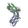
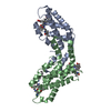
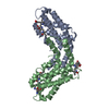
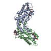
| Entry | Database: PDB / ID: 1xm9 | ||||||
|---|---|---|---|---|---|---|---|
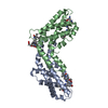
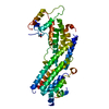
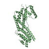
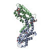
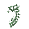
| Title | Structure of the armadillo repeat domain of plakophilin 1 | ||||||
 Components Components | plakophilin 1 | ||||||
 Keywords Keywords | CELL ADHESION / armadillo repeat | ||||||
| Function / homology |  Function and homology information Function and homology informationdesmosome maintenance / intermediate filament bundle assembly / negative regulation of mRNA catabolic process / intermediate filament binding / structural constituent of skin epidermis / positive regulation of cap-dependent translational initiation / Keratinization / ameloblast differentiation / desmosome assembly / transepithelial water transport ...desmosome maintenance / intermediate filament bundle assembly / negative regulation of mRNA catabolic process / intermediate filament binding / structural constituent of skin epidermis / positive regulation of cap-dependent translational initiation / Keratinization / ameloblast differentiation / desmosome assembly / transepithelial water transport / desmosome / positive regulation of cell-cell adhesion / nuclear stress granule / Formation of the cornified envelope / cornified envelope / lamin binding / positive regulation of keratinocyte differentiation / Apoptotic cleavage of cell adhesion proteins / positive regulation of protein localization to membrane / intermediate filament / ficolin-1-rich granule membrane / positive regulation of protein localization to plasma membrane / adherens junction / cell-cell adhesion / cytoplasmic stress granule / cell adhesion / cadherin binding / Neutrophil degranulation / positive regulation of gene expression / perinuclear region of cytoplasm / signal transduction / positive regulation of transcription by RNA polymerase II / DNA binding / RNA binding / nucleoplasm / nucleus / plasma membrane / cytoplasm Similarity search - Function | ||||||
| Biological species |  Homo sapiens (human) Homo sapiens (human) | ||||||
| Method |  X-RAY DIFFRACTION / SYNCHROTRON / SAD / Resolution: 2.8 Å X-RAY DIFFRACTION / SYNCHROTRON / SAD / Resolution: 2.8 Å | ||||||
 Authors Authors | Choi, H.J. / Weis, W.I. | ||||||
 Citation Citation | Journal: J.Mol.Biol. / Year: 2005 Title: Structure of the armadillo repeat domain of plakophilin 1. Authors: Choi, H.J. / Weis, W.I. | ||||||
| History |
|
- Structure visualization
| Structure viewer | Molecule: MolmilJmol/JSmol |
|---|
- Downloads & links
-Download
| PDBx/mmCIF format | 1xm9.cif.gz | 91.1 KB | Display | PDBx/mmCIF format |
|---|---|---|---|---|
| PDB format | pdb1xm9.ent.gz | 69.1 KB | Display | PDB format |
| PDBx/mmJSON format | 1xm9.json.gz | Tree view | PDBx/mmJSON format | |
| Others |  Other downloads Other downloads |
-Validation report
| Arichive directory | https://data.pdbj.org/pub/pdb/validation_reports/xm/1xm9ftp://data.pdbj.org/pub/pdb/validation_reports/xm/1xm9 | HTTPS FTP |
|---|
-Related structure data
| Similar structure data |
|---|
-Links
 PDBj
PDBj


- Assembly
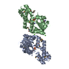
| Deposited unit | 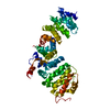
| ||||||||
|---|---|---|---|---|---|---|---|---|---|
| 1 |
| ||||||||
| Unit cell |
|
-Components
| #1: Protein | Mass: 50489.098 Da / Num. of mol.: 1 / Fragment: Armadillo repeat domain Source method: isolated from a genetically manipulated source Source: (gene. exp.) Homo sapiens (human) / Gene: PKP1 / Species (production host): Escherichia coli / Production host:  |
|---|
-Experimental details
-Experiment
| Experiment | Method: X-RAY DIFFRACTION / Number of used crystals: 1 |
|---|
- Sample preparation
| Crystal | Density Matthews: 2.66 Å3/Da / Density % sol: 52 % |
|---|---|
| Crystal grow | Temperature: 293 K / Method: vapor diffusion, hanging drop / pH: 8.6 Details: PEG 3350, Tris-Cl, potassium formate, pH 8.6, VAPOR DIFFUSION, HANGING DROP, temperature 293K |
-Data collection
| Diffraction | Mean temperature: 93 K |
|---|---|
| Diffraction source | Source: SYNCHROTRON / Site: ALS  / Beamline: 8.2.2 / Wavelength: 0.979 Å / Beamline: 8.2.2 / Wavelength: 0.979 Å |
| Detector | Type: ADSC QUANTUM 4 / Detector: CCD / Date: Jul 20, 2003 |
| Radiation | Monochromator: double crystal Si 111 / Protocol: SINGLE WAVELENGTH / Monochromatic (M) / Laue (L): M / Scattering type: x-ray |
| Radiation wavelength | Wavelength: 0.979 Å / Relative weight: 1 |
| Reflection | Resolution: 2.8→48 Å / Num. all: 14705 / Num. obs: 13069 / % possible obs: 89 % / Observed criterion σ(F): 1 / Observed criterion σ(I): 1 / Biso Wilson estimate: 48.7 Å2 / Rsym value: 0.055 |
| Reflection shell | Resolution: 2.8→2.9 Å / % possible all: 73 |
- Processing
| Software |
| ||||||||||||||||||||||||||||||||||||
|---|---|---|---|---|---|---|---|---|---|---|---|---|---|---|---|---|---|---|---|---|---|---|---|---|---|---|---|---|---|---|---|---|---|---|---|---|---|
| Refinement | Method to determine structure: SAD / Resolution: 2.8→29.92 Å / Rfactor Rfree error: 0.01 / Data cutoff high absF: 2496803.59 / Data cutoff low absF: 0 / Isotropic thermal model: RESTRAINED / Cross valid method: THROUGHOUT / σ(F): 0 / Stereochemistry target values: Engh & Huber
| ||||||||||||||||||||||||||||||||||||
| Solvent computation | Solvent model: FLAT MODEL / Bsol: 18.2814 Å2 / ksol: 0.287631 e/Å3 | ||||||||||||||||||||||||||||||||||||
| Displacement parameters | Biso mean: 70.3 Å2
| ||||||||||||||||||||||||||||||||||||
| Refine analyze |
| ||||||||||||||||||||||||||||||||||||
| Refinement step | Cycle: LAST / Resolution: 2.8→29.92 Å
| ||||||||||||||||||||||||||||||||||||
| Refine LS restraints |
| ||||||||||||||||||||||||||||||||||||
| LS refinement shell | Resolution: 2.8→2.98 Å / Rfactor Rfree error: 0.038 / Total num. of bins used: 6
| ||||||||||||||||||||||||||||||||||||
| Xplor file | Serial no: 1 / Param file: PROTEIN_REP.PARAM / Topol file: PROTEIN.TOP |
