 Movie
Movie Controller


+ Open data

- Basic information
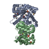
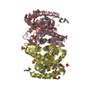
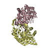
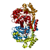
| Entry | Database: PDB / ID: 1xkj | ||||||
|---|---|---|---|---|---|---|---|
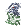
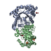
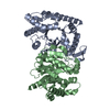
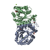
| Title | BACTERIAL LUCIFERASE BETA2 HOMODIMER | ||||||
 Components Components | BETA2 LUCIFERASE | ||||||
 Keywords Keywords | LUMINESCENCE / LUCIFERASE / PHOTOPROTEIN / OXIDOREDUCTASE | ||||||
| Function / homology |  Function and homology information Function and homology informationbacterial luciferase / alkanal monooxygenase (FMN-linked) activity / bioluminescence / cytosol Similarity search - Function | ||||||
| Biological species |  Vibrio harveyi (bacteria) Vibrio harveyi (bacteria) | ||||||
| Method |  X-RAY DIFFRACTION / SYNCHROTRON / MOLECULAR REPLACEMENT / Resolution: 2.5 Å X-RAY DIFFRACTION / SYNCHROTRON / MOLECULAR REPLACEMENT / Resolution: 2.5 Å | ||||||
 Authors Authors | Tanner, J.J. / Krause, K.L. | ||||||
 Citation Citation | Journal: Biochemistry / Year: 1997 Title: Structure of bacterial luciferase beta 2 homodimer: implications for flavin binding. Authors: Tanner, J.J. / Miller, M.D. / Wilson, K.S. / Tu, S.C. / Krause, K.L. | ||||||
| History |
|
- Structure visualization
| Structure viewer | Molecule: MolmilJmol/JSmol |
|---|
- Downloads & links
-Download
| PDBx/mmCIF format | 1xkj.cif.gz | 132.6 KB | Display | PDBx/mmCIF format |
|---|---|---|---|---|
| PDB format | pdb1xkj.ent.gz | 105.5 KB | Display | PDB format |
| PDBx/mmJSON format | 1xkj.json.gz | Tree view | PDBx/mmJSON format | |
| Others |  Other downloads Other downloads |
-Validation report
| Summary document | 1xkj_validation.pdf.gz | 371.2 KB | Display | wwPDB validaton report |
|---|---|---|---|---|
| Full document | 1xkj_full_validation.pdf.gz | 375.9 KB | Display | |
| Data in XML | 1xkj_validation.xml.gz | 13.2 KB | Display | |
| Data in CIF | 1xkj_validation.cif.gz | 20.5 KB | Display | |
| Arichive directory | https://data.pdbj.org/pub/pdb/validation_reports/xk/1xkjftp://data.pdbj.org/pub/pdb/validation_reports/xk/1xkj | HTTPS FTP |
-Related structure data
| Related structure data |  1brlS S: Starting model for refinement |
|---|---|
| Similar structure data |


-Links
 PDBj
PDBj
- Assembly
| Deposited unit | 
| ||||||||
|---|---|---|---|---|---|---|---|---|---|
| 1 |
| ||||||||
| Unit cell |
| ||||||||
| Noncrystallographic symmetry (NCS) | NCS oper: (Code: given Matrix: (0.756074, 0.000164, 0.654486), Vector: |
-Components
| #1: Protein | Mass: 36384.684 Da / Num. of mol.: 2 Source method: isolated from a genetically manipulated source Source: (gene. exp.) Vibrio harveyi (bacteria) / Gene: VIBRIO HARVEYI LUXAB / Plasmid: PTH3 / Gene (production host): VIBRIO HARVEYI LUXAB / Production host: #2: Water | ChemComp-HOH / |  Mass: 18.015 Da / Num. of mol.: 31 / Source method: isolated from a natural source / Formula: H2O Mass: 18.015 Da / Num. of mol.: 31 / Source method: isolated from a natural source / Formula: H2O |
|---|
-Experimental details
-Experiment
| Experiment | Method: X-RAY DIFFRACTION / Number of used crystals: 2 |
|---|
- Sample preparation
| Crystal | Density Matthews: 3.5 Å3/Da / Density % sol: 64 % / Description: DATA MERGED FROM TWO CRYSTALS | |||||||||||||||||||||||||
|---|---|---|---|---|---|---|---|---|---|---|---|---|---|---|---|---|---|---|---|---|---|---|---|---|---|---|
| Crystal grow | Temperature: 277 K / Method: vapor diffusion, sitting drop / pH: 6.2 Details: SITTING DROP, 277 KELVIN, 12% PEG 4000, 17-21% ISOPROPANOL, 0.006M DTT, 0.1M CITRATE PH 6.2. 50 MG/ML PROTEIN., vapor diffusion - sitting drop | |||||||||||||||||||||||||
| Crystal grow | *PLUS Temperature: 4 ℃ / Method: vapor diffusion, sitting drop | |||||||||||||||||||||||||
| Components of the solutions | *PLUS
|
-Data collection
| Diffraction | Mean temperature: 293 K |
|---|---|
| Diffraction source | Source: SYNCHROTRON / Site: EMBL/DESY, HAMBURG  / Beamline: BW7B / Wavelength: 0.865 / Beamline: BW7B / Wavelength: 0.865 |
| Detector | Type: MARRESEARCH / Detector: IMAGE PLATE / Date: Oct 6, 1994 |
| Radiation | Monochromatic (M) / Laue (L): M / Scattering type: x-ray |
| Radiation wavelength | Wavelength: 0.865 Å / Relative weight: 1 |
| Reflection | Resolution: 2.5→15 Å / Num. obs: 32225 / % possible obs: 93 % / Redundancy: 8 % / Biso Wilson estimate: 32.1 Å2 / Rmerge(I) obs: 0.088 |
| Reflection | *PLUS Num. measured all: 245416 |
| Reflection shell | *PLUS Highest resolution: 2.5 Å / Lowest resolution: 2.6 Å / % possible obs: 78 % |
- Processing
| Software |
| ||||||||||||||||||||||||||||||||||||||||||||||||||||||||||||||||||||||||||||||||
|---|---|---|---|---|---|---|---|---|---|---|---|---|---|---|---|---|---|---|---|---|---|---|---|---|---|---|---|---|---|---|---|---|---|---|---|---|---|---|---|---|---|---|---|---|---|---|---|---|---|---|---|---|---|---|---|---|---|---|---|---|---|---|---|---|---|---|---|---|---|---|---|---|---|---|---|---|---|---|---|---|---|
| Refinement | Method to determine structure: MOLECULAR REPLACEMENT Starting model: LUCIFERASE AB HETERODIMER (PDB ENTRY 1BRL) Resolution: 2.5→15 Å / Rfactor Rfree error: 0.004 / Data cutoff high absF: 10000000 / Data cutoff low absF: 0.001 / Isotropic thermal model: RESTRAINED / Cross valid method: THROUGHOUT / σ(F): 0 / Details: BULK SOLVENT MODEL USED
| ||||||||||||||||||||||||||||||||||||||||||||||||||||||||||||||||||||||||||||||||
| Displacement parameters | Biso mean: 37.8 Å2 | ||||||||||||||||||||||||||||||||||||||||||||||||||||||||||||||||||||||||||||||||
| Refine analyze |
| ||||||||||||||||||||||||||||||||||||||||||||||||||||||||||||||||||||||||||||||||
| Refinement step | Cycle: LAST / Resolution: 2.5→15 Å
| ||||||||||||||||||||||||||||||||||||||||||||||||||||||||||||||||||||||||||||||||
| Refine LS restraints |
| ||||||||||||||||||||||||||||||||||||||||||||||||||||||||||||||||||||||||||||||||
| LS refinement shell | Resolution: 2.5→2.66 Å / Rfactor Rfree error: 0.017 / Total num. of bins used: 6
| ||||||||||||||||||||||||||||||||||||||||||||||||||||||||||||||||||||||||||||||||
| Xplor file |
| ||||||||||||||||||||||||||||||||||||||||||||||||||||||||||||||||||||||||||||||||
| Software | *PLUS Name: X-PLOR / Version: 3.843 / Classification: refinement | ||||||||||||||||||||||||||||||||||||||||||||||||||||||||||||||||||||||||||||||||
| Refine LS restraints | *PLUS
|
