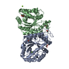
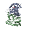
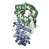
Entry Database : PDB / ID : 5lxeTitle F420-dependent glucose-6-phosphate dehydrogenase from Rhodococcus jostii RHA1 F420-dependent glucose-6-phosphate dehydrogenase 1 Keywords / / / / Function / homology Function Domain/homology Component
/ / / / / / / / / / / / / Biological species Rhodococcus jostii (bacteria)Method / / / Resolution : 1.47 Å Authors Nguyen, Q.-T. / Trinco, G. / Binda, C. / Mattevi, A. / Fraaije, M.W. Funding support Organization Grant number Country The European Community Seventh Framework Programme FP7 (2007--2013) under BioStruct-X (Grants 7551 and 10205) University of Groningen Ubbo Emmius scholarship
Journal : Appl. Microbiol. Biotechnol. / Year : 2017Title : Discovery and characterization of an F420-dependent glucose-6-phosphate dehydrogenase (Rh-FGD1) from Rhodococcus jostii RHA1.Authors : Nguyen, Q.-T. / Trinco, G. / Binda, C. / Mattevi, A. / Fraaije, M.W. History Deposition Sep 20, 2016 Deposition site / Processing site Revision 1.0 Dec 28, 2016 Provider / Type Revision 1.1 Mar 29, 2017 Group Revision 1.2 Jun 7, 2017 Group Revision 1.3 Jan 31, 2018 Group / Category Revision 1.4 Jan 17, 2024 Group / Database references / Refinement descriptionCategory chem_comp_atom / chem_comp_bond ... chem_comp_atom / chem_comp_bond / database_2 / pdbx_initial_refinement_model / struct_ncs_dom_lim Item _database_2.pdbx_DOI / _database_2.pdbx_database_accession ... _database_2.pdbx_DOI / _database_2.pdbx_database_accession / _struct_ncs_dom_lim.beg_auth_comp_id / _struct_ncs_dom_lim.beg_label_asym_id / _struct_ncs_dom_lim.beg_label_comp_id / _struct_ncs_dom_lim.beg_label_seq_id / _struct_ncs_dom_lim.end_auth_comp_id / _struct_ncs_dom_lim.end_label_asym_id / _struct_ncs_dom_lim.end_label_comp_id / _struct_ncs_dom_lim.end_label_seq_id
Show all Show less
 Movie
Movie Controller

 Yorodumi
 Open data
 Basic information
 Components
 Keywords
 Function and homology information
 Rhodococcus jostii (bacteria)
 X-RAY DIFFRACTION /
  Authors
 Italy,
  Netherlands, 2items
  Citation
 Structure visualization
 Downloads & links
 Other downloads


 PDBj
PDBj
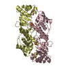
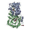
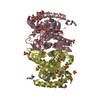
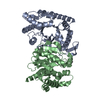
 Assembly
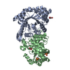

 Mass: 92.094 Da / Num. of mol.: 2 / Source method: obtained synthetically / Formula: C3H8O3
 Mass: 96.063 Da / Num. of mol.: 2 / Source method: obtained synthetically / Formula: SO4
 Mass: 18.015 Da / Num. of mol.: 571 / Source method: isolated from a natural source / Formula: H2O
 Sample preparation
 / Beamline: X06DA / Wavelength: 0.71, 2.25
 Processing