 Movie
Movie Controller

[English] 日本語
 Yorodumi
- PDB-1xjw: The Structure of E. coli Aspartate Transcarbamoylase Q137A Mutant... -
+ Open data

- Basic information
| Entry | Database: PDB / ID: 1xjw | ||||||
|---|---|---|---|---|---|---|---|
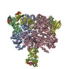
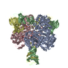
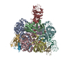
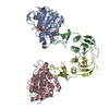
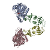
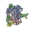
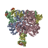
| Title | The Structure of E. coli Aspartate Transcarbamoylase Q137A Mutant in The R-State | ||||||
 Components Components |
| ||||||
 Keywords Keywords | TRANSFERASE/TRANSFERASE REGULATOR / Allosteric enzyme / polar contacts / electrostatics / small angle x-ray scattering / domain closure / intersubunit interactions / TRANSFERASE-TRANSFERASE REGULATOR COMPLEX | ||||||
| Function / homology |  Function and homology information Function and homology informationaspartate carbamoyltransferase complex / pyrimidine nucleotide biosynthetic process / aspartate carbamoyltransferase / aspartate carbamoyltransferase activity / L-glutamine metabolic process / amino acid binding / protein homotrimerization / 'de novo' UMP biosynthetic process / 'de novo' pyrimidine nucleobase biosynthetic process / zinc ion binding ...aspartate carbamoyltransferase complex / pyrimidine nucleotide biosynthetic process / aspartate carbamoyltransferase / aspartate carbamoyltransferase activity / L-glutamine metabolic process / amino acid binding / protein homotrimerization / 'de novo' UMP biosynthetic process / 'de novo' pyrimidine nucleobase biosynthetic process / zinc ion binding / identical protein binding / cytoplasm / cytosol Similarity search - Function | ||||||
| Biological species |  | ||||||
| Method |  X-RAY DIFFRACTION / Refinement from pre-existing model / Resolution: 2.71 Å X-RAY DIFFRACTION / Refinement from pre-existing model / Resolution: 2.71 Å | ||||||
 Authors Authors | Stieglitz, K.A. / Alam, N. / Xia, J. / Gourinath, S. / Tsuruta, H. / Kantrowitz, E.R. | ||||||
 Citation Citation | Journal: J.Mol.Biol. / Year: 2005 Title: A Single Amino Acid Substitution in the Active Site of Escherichia coli Aspartate Transcarbamoylase Prevents the Allosteric Transition. Authors: Stieglitz, K.A. / Pastra-Landis, S.C. / Xia, J. / Tsuruta, H. / Kantrowitz, E.R. | ||||||
| History |
|
- Structure visualization
| Structure viewer | Molecule: MolmilJmol/JSmol |
|---|
- Downloads & links
-Download
| PDBx/mmCIF format | 1xjw.cif.gz | 189.7 KB | Display | PDBx/mmCIF format |
|---|---|---|---|---|
| PDB format | pdb1xjw.ent.gz | 152 KB | Display | PDB format |
| PDBx/mmJSON format | 1xjw.json.gz | Tree view | PDBx/mmJSON format | |
| Others |  Other downloads Other downloads |
-Validation report
| Arichive directory | https://data.pdbj.org/pub/pdb/validation_reports/xj/1xjwftp://data.pdbj.org/pub/pdb/validation_reports/xj/1xjw | HTTPS FTP |
|---|
-Related structure data
| Related structure data |  1d09S S: Starting model for refinement |
|---|---|
| Similar structure data |


-Links
 PDBj
PDBj
- Assembly
| Deposited unit | 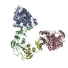
| ||||||||
|---|---|---|---|---|---|---|---|---|---|
| 1 | 
| ||||||||
| Unit cell |
|
-Components
| #1: Protein | Mass: 34280.059 Da / Num. of mol.: 2 / Mutation: Q137A Source method: isolated from a genetically manipulated source Source: (gene. exp.) #2: Protein | Mass: 17143.625 Da / Num. of mol.: 2 Source method: isolated from a genetically manipulated source Source: (gene. exp.) #3: Chemical |   Mass: 255.119 Da / Num. of mol.: 2 / Source method: obtained synthetically / Formula: C6H10NO8P Mass: 255.119 Da / Num. of mol.: 2 / Source method: obtained synthetically / Formula: C6H10NO8P#4: Chemical |   Mass: 65.409 Da / Num. of mol.: 2 / Source method: obtained synthetically / Formula: Zn Mass: 65.409 Da / Num. of mol.: 2 / Source method: obtained synthetically / Formula: Zn#5: Water | ChemComp-HOH / |  Mass: 18.015 Da / Num. of mol.: 116 / Source method: isolated from a natural source / Formula: H2O Mass: 18.015 Da / Num. of mol.: 116 / Source method: isolated from a natural source / Formula: H2O |
|---|
-Experimental details
-Experiment
| Experiment | Method: X-RAY DIFFRACTION / Number of used crystals: 1 |
|---|
- Sample preparation
| Crystal | Density Matthews: 2.5 Å3/Da / Density % sol: 55 % |
|---|---|
| Crystal grow | Temperature: 295 K / Method: microdialysis / pH: 5.9 Details: 12 mg/ml Buffer 50 mM Maleic Acid, 3 mM Sodium Azide 1 mM N-Phosphonacetyl-L-Aspartate, pH 5.9, MICRODIALYSIS, temperature 295K |
-Data collection
| Diffraction | Mean temperature: 298 K |
|---|---|
| Diffraction source | Source: ROTATING ANODE / Type: RIGAKU RU200 / Wavelength: 1.5418 Å |
| Detector | Type: UCSD MARK III / Detector: AREA DETECTOR / Date: Dec 30, 1997 |
| Radiation | Monochromator: GRAPHITE / Protocol: SINGLE WAVELENGTH / Monochromatic (M) / Laue (L): M / Scattering type: x-ray |
| Radiation wavelength | Wavelength: 1.5418 Å / Relative weight: 1 |
| Reflection | Resolution: 2.7→7 Å / Num. all: 40234 / Num. obs: 35809 / % possible obs: 89 % / Observed criterion σ(F): 0 / Observed criterion σ(I): 0 / Redundancy: 2.8 % / Rmerge(I) obs: 0.0875 / Net I/σ(I): 10.5 |
| Reflection shell | Resolution: 2.7→2.8 Å / Redundancy: 2.9 % / Rmerge(I) obs: 0.329 / Mean I/σ(I) obs: 2.59 / Num. unique all: 1380 / % possible all: 85 |
- Processing
| Software |
| |||||||||||||||||||||||||
|---|---|---|---|---|---|---|---|---|---|---|---|---|---|---|---|---|---|---|---|---|---|---|---|---|---|---|
| Refinement | Method to determine structure: Refinement from pre-existing model Starting model: 1D09 Resolution: 2.71→7 Å / Isotropic thermal model: Isotropic / Cross valid method: THROUGHOUT / σ(F): 0 / σ(I): 0 / Stereochemistry target values: Engh & Huber
| |||||||||||||||||||||||||
| Refinement step | Cycle: LAST / Resolution: 2.71→7 Å
|
