 Movie
Movie Controller

[English] 日本語
 Yorodumi
- PDB-1xft: Synchrotron X-ray Powder Diffraction Study of Hexagonal Turkey Eg... -
+ Open data

- Basic information
| Entry | Database: PDB / ID: 1xft | ||||||
|---|---|---|---|---|---|---|---|
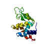
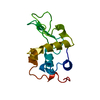
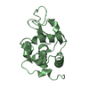
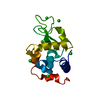
| Title | Synchrotron X-ray Powder Diffraction Study of Hexagonal Turkey Egg-white Lysozyme | ||||||
 Components Components | Lysozyme C | ||||||
 Keywords Keywords | HYDROLASE / powder diffraction / lysozyme / x-rays | ||||||
| Function / homology |  Function and homology information Function and homology informationglycosaminoglycan binding / cell wall macromolecule catabolic process / lysozyme / lysozyme activity / killing of cells of another organism / defense response to Gram-negative bacterium / defense response to Gram-positive bacterium / : / identical protein binding / cytoplasm Similarity search - Function | ||||||
| Biological species |  | ||||||
| Method | POWDER DIFFRACTION /  SYNCHROTRON / MOLECULAR REPLACEMENT / Resolution: 3.35 Å SYNCHROTRON / MOLECULAR REPLACEMENT / Resolution: 3.35 Å | ||||||
 Authors Authors | Margiolaki, I. / Wright, J.P. | ||||||
 Citation Citation | Journal: Acta Crystallogr.,Sect.D / Year: 2005 Title: Synchrotron X-ray powder diffraction study of hexagonal turkey egg-white lysozyme. Authors: Margiolaki, I. / Wright, J.P. / Fitch, A.N. / Fox, G.C. / Von Dreele, R.B. #1: Journal: Acta Crystallogr.,Sect.D / Year: 2001Title: Binding of N-acetylglucosamine to chicken egg lysozyme: a powder diffraction study Authors: Von Dreele, R.B. #2: Journal: Acta Crystallogr.,Sect.D / Year: 1995Title: Structure of hexagonal turkey egg-white lysozyme at 1.65A resolution Authors: Howell, P.L. #3: Journal: Acta Crystallogr.,Sect.D / Year: 1993Title: X-ray structure of monoclinic turkey egg lysozyme at 1.3 A resolution Authors: Harata, K. #4: Journal: Acta Crystallogr.,Sect.B / Year: 1992Title: Structure determination of turkey egg-white lysozyme using Laue diffraction data Authors: Howell, P.L. / Almo, S.C. / Parsons, M.R. / Hajdu, J. / Petsko, G.A. | ||||||
| History |
|
- Structure visualization
| Structure viewer | Molecule: MolmilJmol/JSmol |
|---|
- Downloads & links
-Download
| PDBx/mmCIF format | 1xft.cif.gz | 33.1 KB | Display | PDBx/mmCIF format |
|---|---|---|---|---|
| PDB format | pdb1xft.ent.gz | 19.9 KB | Display | PDB format |
| PDBx/mmJSON format | 1xft.json.gz | Tree view | PDBx/mmJSON format | |
| Others |  Other downloads Other downloads |
-Validation report
| Arichive directory | https://data.pdbj.org/pub/pdb/validation_reports/xf/1xftftp://data.pdbj.org/pub/pdb/validation_reports/xf/1xft | HTTPS FTP |
|---|
-Related structure data
| Related structure data |  1tewS S: Starting model for refinement |
|---|---|
| Similar structure data |

-Links
 PDBj
PDBj
- Assembly
| Deposited unit | 
|
|---|---|
| 1 |
|
-Components
| #1: Protein | Mass: 14228.105 Da / Num. of mol.: 1 / Fragment: LYSOZYME / Source method: isolated from a natural source / Details: sigma-aldrich chemical company LOT. 064H7230 / Source: (natural) |
|---|---|
| Has protein modification | Y |
-Experimental details
-Experiment
| Experiment | Method: POWDER DIFFRACTION |
|---|
- Sample preparation
| Crystal | Density Matthews: 2.25 Å3/Da / Density % sol: 44.81 % |
|---|---|
| Crystal grow | Temperature: 295 K / Method: salting out / pH: 6 / Details: NaCl, pH 6, SALTING OUT, temperature 295K |
-Data collection
| Diffraction | Mean temperature: 295 K |
|---|---|
| Diffraction source | Source: SYNCHROTRON / Site: ESRF  / Beamline: ID31 / Wavelength: 0.700667 Å / Beamline: ID31 / Wavelength: 0.700667 Å |
| Detector | Type: CUSTOM-MADE / Detector: DIFFRACTOMETER / Date: Dec 3, 2003 Details: DOUBLE SI(111) MONOCHROMATOR, 9 CHANNEL SI(111) MULTI-ANALYSER |
| Radiation | Monochromator: DOUBLE SI(111) / Protocol: SINGLE WAVELENGTH / Monochromatic (M) / Laue (L): M / Scattering type: x-ray |
| Radiation wavelength | Wavelength: 0.700667 Å / Relative weight: 1 |
| Reflection | Resolution: 3.35→95.1 Å / Num. all: 2099 / Num. obs: 2099 / Observed criterion σ(F): 0 / Observed criterion σ(I): 0 |
- Processing
| Software |
| ||||||||||||||||||||||||||||||||||||||||||||||||||||||||||||
|---|---|---|---|---|---|---|---|---|---|---|---|---|---|---|---|---|---|---|---|---|---|---|---|---|---|---|---|---|---|---|---|---|---|---|---|---|---|---|---|---|---|---|---|---|---|---|---|---|---|---|---|---|---|---|---|---|---|---|---|---|---|
| Refinement | Method to determine structure: MOLECULAR REPLACEMENT Starting model: 1TEW Resolution: 3.35→95.1 Å / Isotropic thermal model: OVERALL / σ(F): 0 / σ(I): 0 / Stereochemistry target values: Engh & Huber / Details: POWDER DIFFRACTION
| ||||||||||||||||||||||||||||||||||||||||||||||||||||||||||||
| Displacement parameters | Biso mean: 23.69 Å2 | ||||||||||||||||||||||||||||||||||||||||||||||||||||||||||||
| Refinement step | Cycle: LAST / Resolution: 3.35→95.1 Å
| ||||||||||||||||||||||||||||||||||||||||||||||||||||||||||||
| Refine LS restraints |
|
