 Movie
Movie Controller

[English] 日本語
 Yorodumi
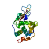
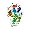
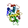
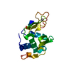
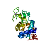
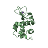
- PDB-1tew: STRUCTURE OF HEXAGONAL TURKEY EGG WHITE LYSOZYME AT 1.65 ANGSTROM... -
+ Open data

- Basic information
| Entry | Database: PDB / ID: 1tew | ||||||
|---|---|---|---|---|---|---|---|
| Title | STRUCTURE OF HEXAGONAL TURKEY EGG WHITE LYSOZYME AT 1.65 ANGSTROMS RESOLUTION | ||||||
 Components Components | TURKEY EGG WHITE LYSOZYME | ||||||
 Keywords Keywords | HYDROLASE(O-GLYCOSYL) | ||||||
| Function / homology |  Function and homology information Function and homology informationglycosaminoglycan binding / cell wall macromolecule catabolic process / lysozyme / lysozyme activity / killing of cells of another organism / defense response to Gram-negative bacterium / defense response to Gram-positive bacterium / : / identical protein binding / cytoplasm Similarity search - Function | ||||||
| Biological species |  | ||||||
| Method |  X-RAY DIFFRACTION / Resolution: 1.65 Å X-RAY DIFFRACTION / Resolution: 1.65 Å | ||||||
 Authors Authors | Howell, P.L. | ||||||
 Citation Citation | Journal: Acta Crystallogr.,Sect.D / Year: 1995 Title: Structure of hexagonal turkey egg-white lysozyme at 1.65A resolution. Authors: Howell, P.L. #1: Journal: Acta Crystallogr.,Sect.D / Year: 1993Title: X-Ray Structure of Monoclinic Turkey Egg White Lysozyme at 1.3 Angstroms Resolution Authors: Harata, K. #2: Journal: Acta Crystallogr.,Sect.B / Year: 1992Title: Structure Determination of Turkey Egg White Lysozyme Using Laue Diffraction Data Authors: Howell, P.L. / Almo, S.C. / Parsons, M.R. / Hajdu, J. / Petsko, G.A. | ||||||
| History |
|
- Structure visualization
| Structure viewer | Molecule: MolmilJmol/JSmol |
|---|
- Downloads & links
-Download
| PDBx/mmCIF format | 1tew.cif.gz | 37.8 KB | Display | PDBx/mmCIF format |
|---|---|---|---|---|
| PDB format | pdb1tew.ent.gz | 25.8 KB | Display | PDB format |
| PDBx/mmJSON format | 1tew.json.gz | Tree view | PDBx/mmJSON format | |
| Others |  Other downloads Other downloads |
-Validation report
| Arichive directory | https://data.pdbj.org/pub/pdb/validation_reports/te/1tewftp://data.pdbj.org/pub/pdb/validation_reports/te/1tew | HTTPS FTP |
|---|
-Related structure data
| Similar structure data |
|---|
-Links
 PDBj
PDBj
- Assembly
| Deposited unit | 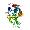
| ||||||||
|---|---|---|---|---|---|---|---|---|---|
| 1 |
| ||||||||
| Unit cell |
| ||||||||
| Components on special symmetry positions |
|
-Components
| #1: Protein | Mass: 14228.105 Da / Num. of mol.: 1 Source method: isolated from a genetically manipulated source Source: (gene. exp.) |
|---|---|
| #2: Chemical | ChemComp-SCN /   Mass: 58.082 Da / Num. of mol.: 1 / Source method: obtained synthetically / Formula: CNS Mass: 58.082 Da / Num. of mol.: 1 / Source method: obtained synthetically / Formula: CNS |
| #3: Water | ChemComp-HOH /  Mass: 18.015 Da / Num. of mol.: 86 / Source method: isolated from a natural source / Formula: H2O Mass: 18.015 Da / Num. of mol.: 86 / Source method: isolated from a natural source / Formula: H2O |
| Has protein modification | Y |
-Experimental details
-Experiment
| Experiment | Method: X-RAY DIFFRACTION |
|---|
- Sample preparation
| Crystal | Density Matthews: 2.12 Å3/Da / Density % sol: 41.98 % | |||||||||||||||||||||||||
|---|---|---|---|---|---|---|---|---|---|---|---|---|---|---|---|---|---|---|---|---|---|---|---|---|---|---|
| Crystal grow | *PLUS pH: 4.5 / Method: vapor diffusion, hanging drop | |||||||||||||||||||||||||
| Components of the solutions | *PLUS
|
-Data collection
| Reflection | Num. obs: 15265 / % possible obs: 97 % / Observed criterion σ(I): 0 |
|---|---|
| Reflection | *PLUS Highest resolution: 1.65 Å / Lowest resolution: 10 Å / Num. measured all: 27701 / Rmerge(I) obs: 0.053 |
| Reflection shell | *PLUS Highest resolution: 1.65 Å / Lowest resolution: 1.7 Å / % possible obs: 80 % |
- Processing
| Software |
| ||||||||||||||||||||||||||||||||||||||||||||||||||||||||||||||||||||||||||||||||
|---|---|---|---|---|---|---|---|---|---|---|---|---|---|---|---|---|---|---|---|---|---|---|---|---|---|---|---|---|---|---|---|---|---|---|---|---|---|---|---|---|---|---|---|---|---|---|---|---|---|---|---|---|---|---|---|---|---|---|---|---|---|---|---|---|---|---|---|---|---|---|---|---|---|---|---|---|---|---|---|---|---|
| Refinement | Resolution: 1.65→8 Å / σ(F): 0
| ||||||||||||||||||||||||||||||||||||||||||||||||||||||||||||||||||||||||||||||||
| Displacement parameters | Biso mean: 23.29 Å2 | ||||||||||||||||||||||||||||||||||||||||||||||||||||||||||||||||||||||||||||||||
| Refine analyze | Luzzati coordinate error obs: 0.15 Å | ||||||||||||||||||||||||||||||||||||||||||||||||||||||||||||||||||||||||||||||||
| Refinement step | Cycle: LAST / Resolution: 1.65→8 Å
| ||||||||||||||||||||||||||||||||||||||||||||||||||||||||||||||||||||||||||||||||
| Refine LS restraints |
| ||||||||||||||||||||||||||||||||||||||||||||||||||||||||||||||||||||||||||||||||
| Software | *PLUS Name: X-PLOR / Classification: refinement | ||||||||||||||||||||||||||||||||||||||||||||||||||||||||||||||||||||||||||||||||
| Refinement | *PLUS Rfactor obs: 0.206 / Rfactor Rwork: 0.206 | ||||||||||||||||||||||||||||||||||||||||||||||||||||||||||||||||||||||||||||||||
| Solvent computation | *PLUS | ||||||||||||||||||||||||||||||||||||||||||||||||||||||||||||||||||||||||||||||||
| Displacement parameters | *PLUS | ||||||||||||||||||||||||||||||||||||||||||||||||||||||||||||||||||||||||||||||||
| Refine LS restraints | *PLUS
|
