 Movie
Movie Controller

[English] 日本語
 Yorodumi
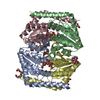
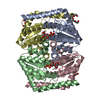
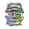
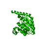
- PDB-1x92: CRYSTAL STRUCTURE OF PSEUDOMONAS AERUGINOSA PHOSPHOHEPTOSE ISOMER... -
+ Open data

- Basic information
| Entry | Database: PDB / ID: 1x92 | ||||||
|---|---|---|---|---|---|---|---|
| Title | CRYSTAL STRUCTURE OF PSEUDOMONAS AERUGINOSA PHOSPHOHEPTOSE ISOMERASE IN COMPLEX WITH REACTION PRODUCT D-GLYCERO-D-MANNOPYRANOSE-7-PHOSPHATE | ||||||
 Components Components | PHOSPHOHEPTOSE ISOMERASE | ||||||
 Keywords Keywords | ISOMERASE / MIDWEST CENTRE FOR STRUCTURAL GENOMICS / SIS Domain / a/b protein / lipopolysaccharide biosynthesis / PSI / Protein Structure Initiative / Midwest Center for Structural Genomics / MCSG | ||||||
| Function / homology |  Function and homology information Function and homology informationDnaA-DiaA complex / D-sedoheptulose-7-phosphate isomerase / D-sedoheptulose 7-phosphate isomerase activity / D-glycero-D-manno-heptose 7-phosphate biosynthetic process / lipopolysaccharide core region biosynthetic process / carbohydrate derivative binding / positive regulation of DNA-templated DNA replication initiation / zinc ion binding / cytoplasm Similarity search - Function | ||||||
| Biological species |   Pseudomonas aeruginosa (bacteria) Pseudomonas aeruginosa (bacteria) | ||||||
| Method |  X-RAY DIFFRACTION / SYNCHROTRON / MOLECULAR REPLACEMENT / Resolution: 2.3 Å X-RAY DIFFRACTION / SYNCHROTRON / MOLECULAR REPLACEMENT / Resolution: 2.3 Å | ||||||
 Authors Authors | Walker, J.R. / Evdokimova, E. / Kudritska, M. / Joachimiak, A. / Edwards, A. / Savchenko, A. / Midwest Center for Structural Genomics (MCSG) | ||||||
 Citation Citation | Journal: J.Biol.Chem. / Year: 2008 Title: Structure and function of sedoheptulose-7-phosphate isomerase, a critical enzyme for lipopolysaccharide biosynthesis and a target for antibiotic adjuvants. Authors: Taylor, P.L. / Blakely, K.M. / de Leon, G.P. / Walker, J.R. / McArthur, F. / Evdokimova, E. / Zhang, K. / Valvano, M.A. / Wright, G.D. / Junop, M.S. #1: Journal: Microbiology / Year: 2002Title: Novel pathways for biosynthesis of nucleotide-activated glycero-manno-heptose precursors of bacterial glycoproteins and cell surface polysaccharides Authors: Valvano, M.A. / Messner, P. / Kosma, P. #2: Journal: J.Biol.Chem. / Year: 1996Title: Biosynthesis of inner core lipopolysaccharide in enteric bacteria identification and characterization of a conserved phosphoheptose isomerase Authors: Brooke, J.S. / Valvano, M.A. | ||||||
| History |
|
- Structure visualization
| Structure viewer | Molecule: MolmilJmol/JSmol |
|---|
- Downloads & links
-Download
| PDBx/mmCIF format | 1x92.cif.gz | 89.2 KB | Display | PDBx/mmCIF format |
|---|---|---|---|---|
| PDB format | pdb1x92.ent.gz | 69.1 KB | Display | PDB format |
| PDBx/mmJSON format | 1x92.json.gz | Tree view | PDBx/mmJSON format | |
| Others |  Other downloads Other downloads |
-Validation report
| Arichive directory | https://data.pdbj.org/pub/pdb/validation_reports/x9/1x92ftp://data.pdbj.org/pub/pdb/validation_reports/x9/1x92 | HTTPS FTP |
|---|
-Related structure data
| Related structure data | 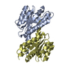 2i22C 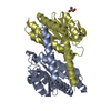 2i2wC 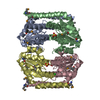 3bjzC C: citing same article ( |
|---|---|
| Similar structure data | |
| Other databases |

-Links
 PDBj
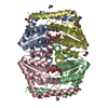
PDBj- Assembly
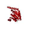
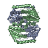
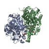
| Deposited unit | 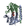
| ||||||||
|---|---|---|---|---|---|---|---|---|---|
| 1 | 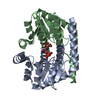
| ||||||||
| Unit cell |
| ||||||||
| Details | The second part of the assembly can be generated by apply the matrix: 0.5000 0.8660 0.0000 0.8660 -.5000 0.0000 0.0000 0.0000 -1.000 63.1685 -109.4127 -18.8724 |
-Components
| #1: Protein | Mass: 21628.498 Da / Num. of mol.: 2 Source method: isolated from a genetically manipulated source Source: (gene. exp.) Pseudomonas aeruginosa (bacteria) / Gene: lpcA, gmhA / Plasmid: modified pET15B / Production host: #2: Sugar |   Type: D-saccharide, alpha linking / Mass: 290.162 Da / Num. of mol.: 2 / Source method: obtained synthetically / Formula: C7H15O10P Type: D-saccharide, alpha linking / Mass: 290.162 Da / Num. of mol.: 2 / Source method: obtained synthetically / Formula: C7H15O10P#3: Water | ChemComp-HOH / |  Mass: 18.015 Da / Num. of mol.: 196 / Source method: isolated from a natural source / Formula: H2O Mass: 18.015 Da / Num. of mol.: 196 / Source method: isolated from a natural source / Formula: H2O |
|---|
-Experimental details
-Experiment
| Experiment | Method: X-RAY DIFFRACTION / Number of used crystals: 1 |
|---|
- Sample preparation
| Crystal | Density Matthews: 3.01 Å3/Da / Density % sol: 59.17 % |
|---|---|
| Crystal grow | Temperature: 298 K / Method: vapor diffusion, hanging drop / pH: 5.6 Details: D-SEDOHEPTULOSE-7-PHOSPHATE, AMMONIUM SULPHATE, K/NA TARTRATE, NA CITRATE, pH 5.6, VAPOR DIFFUSION, HANGING DROP, temperature 298.0K |
-Data collection
| Diffraction | Mean temperature: 100 K |
|---|---|
| Diffraction source | Source: SYNCHROTRON / Site: APS  / Beamline: 17-ID / Wavelength: 1.07229 Å / Beamline: 17-ID / Wavelength: 1.07229 Å |
| Detector | Type: ADSC QUANTUM 210 / Detector: CCD / Date: Aug 6, 2004 / Details: MIRRORS |
| Radiation | Monochromator: SI(111)DOUBLE CRYSTAL / Protocol: SINGLE WAVELENGTH / Monochromatic (M) / Laue (L): M / Scattering type: x-ray |
| Radiation wavelength | Wavelength: 1.07229 Å / Relative weight: 1 |
| Reflection | Resolution: 2.3→40 Å / Num. all: 24669 / Num. obs: 24669 / % possible obs: 99.9 % / Observed criterion σ(F): 0 / Observed criterion σ(I): -3 / Redundancy: 12.4 % / Biso Wilson estimate: 21.3 Å2 / Rmerge(I) obs: 0.125 / Net I/σ(I): 25.03 |
| Reflection shell | Resolution: 2.3→2.38 Å / Redundancy: 12.6 % / Rmerge(I) obs: 0.404 / Mean I/σ(I) obs: 6.17 / % possible all: 100 |
- Processing
| Software |
| ||||||||||||||||||||||||||||||||||||
|---|---|---|---|---|---|---|---|---|---|---|---|---|---|---|---|---|---|---|---|---|---|---|---|---|---|---|---|---|---|---|---|---|---|---|---|---|---|
| Refinement | Method to determine structure: MOLECULAR REPLACEMENT Starting model: STRUCTURE DETERMINED BY MAD PHASING IN SPACEGROUP P21212 Resolution: 2.3→39.34 Å / Rfactor Rfree error: 0.006 / Data cutoff high absF: 2787370.22 / Data cutoff low absF: 0 / Isotropic thermal model: RESTRAINED / Cross valid method: THROUGHOUT / σ(F): 0 / Stereochemistry target values: Engh & Huber
| ||||||||||||||||||||||||||||||||||||
| Solvent computation | Solvent model: FLAT MODEL / Bsol: 29.4621 Å2 / ksol: 0.353441 e/Å3 | ||||||||||||||||||||||||||||||||||||
| Displacement parameters | Biso mean: 29.1 Å2
| ||||||||||||||||||||||||||||||||||||
| Refine analyze |
| ||||||||||||||||||||||||||||||||||||
| Refinement step | Cycle: LAST / Resolution: 2.3→39.34 Å
| ||||||||||||||||||||||||||||||||||||
| Refine LS restraints |
| ||||||||||||||||||||||||||||||||||||
| LS refinement shell | Resolution: 2.3→2.44 Å / Rfactor Rfree error: 0.02 / Total num. of bins used: 6
| ||||||||||||||||||||||||||||||||||||
| Xplor file |
|
