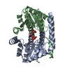
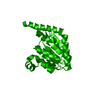
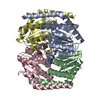
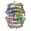
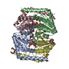
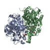
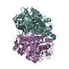
- PDB-4u6n: Crystal structure of Escherichia coli DiaA -
+
Open data
ID or keywords:
Loading...
-
Basic information
Entry
Database: PDB / ID: 4u6n
Title
Crystal structure of Escherichia coli DiaA
Components
DnaA initiator-associating protein DiaA
Keywords
REPLICATION / DiaA / DnaA initiator-associating protein / DNA replication / SIS family
Function / homology
Function and homology information
DnaA-DiaA complex / carbohydrate derivative metabolic process / carbohydrate derivative binding / positive regulation of DNA-templated DNA replication initiation / DNA replication / protein homodimerization activity / identical protein binding Similarity search - Function
Resolution: 1.91→60.65 Å / Cor.coef. Fo:Fc: 0.972 / Cor.coef. Fo:Fc free: 0.966 / SU B: 9.425 / SU ML: 0.129 / Cross valid method: THROUGHOUT / ESU R: 0.137 / ESU R Free: 0.125 / Stereochemistry target values: MAXIMUM LIKELIHOOD / Details: HYDROGENS HAVE BEEN ADDED IN THE RIDING POSITIONS
Rfactor
Num. reflection
% reflection
Selection details
Rfree
0.19841
750
5 %
RANDOM
Rwork
0.16489
-
-
-
obs
0.1665
14188
99.86 %
-
Solvent computation
Ion probe radii: 0.8 Å / Shrinkage radii: 0.8 Å / VDW probe radii: 1.2 Å / Solvent model: MASK
 Movie
Movie Controller


 Open data
 Basic information
 Components
 Keywords
 Function and homology information
 X-RAY DIFFRACTION /
  Authors
 Australia, 1items
  Citation
 Structure visualization
 Downloads & links
 Other downloads
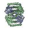


 PDBj
PDBj Assembly
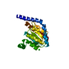

 Mass: 35.453 Da / Num. of mol.: 1 / Source method: obtained synthetically / Formula: Cl
 Mass: 18.015 Da / Num. of mol.: 48 / Source method: isolated from a natural source / Formula: H2O
 Sample preparation
 Processing