 Movie
Movie Controller

[English] 日本語
 Yorodumi
- PDB-1x91: Crystal structure of mutant form A of a pectin methylesterase inh... -
+ Open data

- Basic information
| Entry | Database: PDB / ID: 1x91 | ||||||
|---|---|---|---|---|---|---|---|
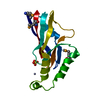
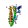
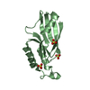
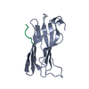
| Title | Crystal structure of mutant form A of a pectin methylesterase inhibitor from Arabidopsis | ||||||
 Components Components | invertase/pectin methylesterase inhibitor family protein | ||||||
 Keywords Keywords | PROTEIN BINDING / four-helix bundle / alpha hairpin / disulfide bridge / domain-swapping / linker / proline / mutant | ||||||
| Function / homology |  Function and homology information Function and homology informationpectinesterase inhibitor activity / pollen tube tip / pollen tube growth / apoplast / extracellular region Similarity search - Function | ||||||
| Biological species |  | ||||||
| Method |  X-RAY DIFFRACTION / SYNCHROTRON / MOLECULAR REPLACEMENT / Resolution: 1.5 Å X-RAY DIFFRACTION / SYNCHROTRON / MOLECULAR REPLACEMENT / Resolution: 1.5 Å | ||||||
 Authors Authors | Hothorn, M. / Wolf, S. / Aloy, P. / Greiner, S. / Scheffzek, K. | ||||||
 Citation Citation | Journal: Plant Cell / Year: 2004 Title: Structural insights into the target specificity of plant invertase and pectin methylesterase inhibitory proteins Authors: Hothorn, M. / Wolf, S. / Aloy, P. / Greiner, S. / Scheffzek, K. | ||||||
| History |
|
- Structure visualization
| Structure viewer | Molecule: MolmilJmol/JSmol |
|---|
- Downloads & links
-Download
| PDBx/mmCIF format | 1x91.cif.gz | 43.8 KB | Display | PDBx/mmCIF format |
|---|---|---|---|---|
| PDB format | pdb1x91.ent.gz | 30.8 KB | Display | PDB format |
| PDBx/mmJSON format | 1x91.json.gz | Tree view | PDBx/mmJSON format | |
| Others |  Other downloads Other downloads |
-Validation report
| Arichive directory | https://data.pdbj.org/pub/pdb/validation_reports/x9/1x91ftp://data.pdbj.org/pub/pdb/validation_reports/x9/1x91 | HTTPS FTP |
|---|
-Related structure data
| Related structure data |  1x8zC  1x90SC C: citing same article ( S: Starting model for refinement |
|---|---|
| Similar structure data |


-Links
 PDBj
PDBj- Assembly
| Deposited unit | 
| |||||||||
|---|---|---|---|---|---|---|---|---|---|---|
| 1 |
| |||||||||
| Unit cell |
| |||||||||
| Components on special symmetry positions |
| |||||||||
| Details | The biologically assembly is consistent with the content of the asymmetric unit, monomer |
-Components
| #1: Protein | Mass: 16418.689 Da / Num. of mol.: 1 / Fragment: residues 1-149 / Mutation: P28A Source method: isolated from a genetically manipulated source Source: (gene. exp.)  |
|---|---|
| #2: Water | ChemComp-HOH /  Mass: 18.015 Da / Num. of mol.: 104 / Source method: isolated from a natural source / Formula: H2O Mass: 18.015 Da / Num. of mol.: 104 / Source method: isolated from a natural source / Formula: H2O |
| Has protein modification | Y |
-Experimental details
-Experiment
| Experiment | Method: X-RAY DIFFRACTION / Number of used crystals: 1 |
|---|
- Sample preparation
| Crystal | Density Matthews: 2.4 Å3/Da / Density % sol: 49 % |
|---|---|
| Crystal grow | Temperature: 298 K / Method: vapor diffusion, hanging drop / pH: 5.6 Details: 0.2M (NH4)2SO4, 0.3M Na/K tartrate, 0.1M Na citrate, pH 5.6, VAPOR DIFFUSION, HANGING DROP, temperature 298K |
-Data collection
| Diffraction | Mean temperature: 100 K |
|---|---|
| Diffraction source | Source: SYNCHROTRON / Site: ESRF  / Beamline: ID14-2 / Wavelength: 0.933 Å / Beamline: ID14-2 / Wavelength: 0.933 Å |
| Detector | Type: ADSC QUANTUM 4 / Detector: CCD / Date: Apr 18, 2004 / Details: toroidal mirror |
| Radiation | Monochromator: Diamond (111), Ge(220) / Protocol: SINGLE WAVELENGTH / Monochromatic (M) / Laue (L): M / Scattering type: x-ray |
| Radiation wavelength | Wavelength: 0.933 Å / Relative weight: 1 |
| Reflection | Resolution: 1.5→54.23 Å / Num. all: 24884 / Num. obs: 24884 / % possible obs: 99.8 % / Observed criterion σ(F): -3 / Observed criterion σ(I): -3 / Redundancy: 3.6 % / Rmerge(I) obs: 0.053 / Net I/σ(I): 15.4 |
| Reflection shell | Resolution: 1.5→1.54 Å / Redundancy: 3.3 % / Rmerge(I) obs: 0.358 / Mean I/σ(I) obs: 3.6 / Num. unique all: 1833 / % possible all: 98.7 |
- Processing
| Software |
| |||||||||||||||||||||||||
|---|---|---|---|---|---|---|---|---|---|---|---|---|---|---|---|---|---|---|---|---|---|---|---|---|---|---|
| Refinement | Method to determine structure: MOLECULAR REPLACEMENT Starting model: PDB ENTRY 1X90 CHAIN A Resolution: 1.5→54.23 Å / Isotropic thermal model: ISOTROPIC / Cross valid method: TROUGHOUT / σ(F): 0 / Stereochemistry target values: Engh & Huber
| |||||||||||||||||||||||||
| Displacement parameters | Biso mean: 15.278 Å2 | |||||||||||||||||||||||||
| Refinement step | Cycle: LAST / Resolution: 1.5→54.23 Å
| |||||||||||||||||||||||||
| Refine LS restraints |
| |||||||||||||||||||||||||
| LS refinement shell | Resolution: 1.5→1.54 Å
|
