 Movie
Movie Controller

[English] 日本語
 Yorodumi
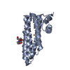
- PDB-1x8z: Crystal structure of a pectin methylesterase inhibitor from Arabi... -
+ Open data

- Basic information
| Entry | Database: PDB / ID: 1x8z | ||||||
|---|---|---|---|---|---|---|---|
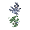
| Title | Crystal structure of a pectin methylesterase inhibitor from Arabidopsis thaliana | ||||||
 Components Components | invertase/pectin methylesterase inhibitor family protein | ||||||
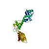
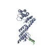
 Keywords Keywords | PROTEIN BINDING / four-helix bundle / alpha hairpin / disulfide bridge | ||||||
| Function / homology |  Function and homology information Function and homology informationpectinesterase inhibitor activity / pollen tube tip / pollen tube growth / apoplast Similarity search - Function | ||||||
| Biological species |  | ||||||
| Method |  X-RAY DIFFRACTION / SYNCHROTRON / MOLECULAR REPLACEMENT / Resolution: 2.86 Å X-RAY DIFFRACTION / SYNCHROTRON / MOLECULAR REPLACEMENT / Resolution: 2.86 Å | ||||||
 Authors Authors | Hothorn, M. / Wolf, S. / Aloy, P. / Greiner, S. / Scheffzek, K. | ||||||
 Citation Citation | Journal: Plant Cell / Year: 2004 Title: Structural insights into the target specificity of plant invertase and pectin methylesterase inhibitory proteins Authors: Hothorn, M. / Wolf, S. / Aloy, P. / Greiner, S. / Scheffzek, K. | ||||||
| History |
|
- Structure visualization
| Structure viewer | Molecule: MolmilJmol/JSmol |
|---|
- Downloads & links
-Download
| PDBx/mmCIF format | 1x8z.cif.gz | 93.7 KB | Display | PDBx/mmCIF format |
|---|---|---|---|---|
| PDB format | pdb1x8z.ent.gz | 73.3 KB | Display | PDB format |
| PDBx/mmJSON format | 1x8z.json.gz | Tree view | PDBx/mmJSON format | |
| Others |  Other downloads Other downloads |
-Validation report
| Arichive directory | https://data.pdbj.org/pub/pdb/validation_reports/x8/1x8zftp://data.pdbj.org/pub/pdb/validation_reports/x8/1x8z | HTTPS FTP |
|---|
-Related structure data
| Related structure data | 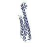 1x90C  1x91C 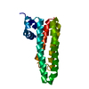 1rj1S S: Starting model for refinement C: citing same article ( |
|---|---|
| Similar structure data |

-Links
 PDBj
PDBj- Assembly
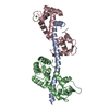
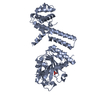
| Deposited unit | 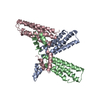
| ||||||||
|---|---|---|---|---|---|---|---|---|---|
| 1 | 
| ||||||||
| 2 | 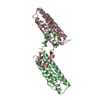
| ||||||||
| 3 |
| ||||||||
| Unit cell |
| ||||||||
| Details | The biological assembly is a dimer generated from the molecules chain B and C in the asymmetric unit / The biological assembly is a dimer generated from the molecule chain A and its closest symmetry mate |
-Components
| #1: Protein | Mass: 16444.725 Da / Num. of mol.: 3 / Fragment: residues 1-149 Source method: isolated from a genetically manipulated source Source: (gene. exp.)  #2: Water | ChemComp-HOH / |  Mass: 18.015 Da / Num. of mol.: 22 / Source method: isolated from a natural source / Formula: H2O Mass: 18.015 Da / Num. of mol.: 22 / Source method: isolated from a natural source / Formula: H2OHas protein modification | Y | |
|---|
-Experimental details
-Experiment
| Experiment | Method: X-RAY DIFFRACTION / Number of used crystals: 1 |
|---|
- Sample preparation
| Crystal | Density Matthews: 3.05 Å3/Da / Density % sol: 60 % |
|---|---|
| Crystal grow | Temperature: 298 K / Method: vapor diffusion, hanging drop / pH: 6.2 Details: 10%(v/v) PEG 8000, 0.3M NaCl, 0.1M Na/K Pi, pH 6.2, VAPOR DIFFUSION, HANGING DROP, temperature 298.0K |
-Data collection
| Diffraction | Mean temperature: 100 K |
|---|---|
| Diffraction source | Source: SYNCHROTRON / Site: SLS  / Beamline: X06SA / Wavelength: 1 Å / Beamline: X06SA / Wavelength: 1 Å |
| Detector | Type: MARRESEARCH / Detector: CCD / Date: Nov 10, 2003 / Details: Dynamically bendable mirror |
| Radiation | Monochromator: LN2 cooled fixed-exit Si(111) monochromator / Protocol: SINGLE WAVELENGTH / Monochromatic (M) / Laue (L): M / Scattering type: x-ray |
| Radiation wavelength | Wavelength: 1 Å / Relative weight: 1 |
| Reflection | Resolution: 2.86→29.06 Å / Num. all: 14136 / Num. obs: 14136 / % possible obs: 98.4 % / Observed criterion σ(F): -3 / Observed criterion σ(I): -3 / Redundancy: 3.5 % / Rmerge(I) obs: 0.059 / Net I/σ(I): 16.91 |
| Reflection shell | Resolution: 2.86→3.04 Å / Redundancy: 3.6 % / Rmerge(I) obs: 0.274 / Mean I/σ(I) obs: 5.4 / Num. unique all: 2307 / % possible all: 98.1 |
- Processing
| Software |
| |||||||||||||||||||||||||
|---|---|---|---|---|---|---|---|---|---|---|---|---|---|---|---|---|---|---|---|---|---|---|---|---|---|---|
| Refinement | Method to determine structure: MOLECULAR REPLACEMENT Starting model: p(ALA) version of PDB entry 1RJ1 Resolution: 2.86→29.06 Å / Isotropic thermal model: ISOTROPIC RESTRAINED / Cross valid method: THROUGHOUT / σ(F): 0 / Stereochemistry target values: Engh & Huber
| |||||||||||||||||||||||||
| Refine analyze |
| |||||||||||||||||||||||||
| Refinement step | Cycle: LAST / Resolution: 2.86→29.06 Å
| |||||||||||||||||||||||||
| Refine LS restraints |
| |||||||||||||||||||||||||
| LS refinement shell | Resolution: 2.86→3.04 Å / Rfactor Rfree error: 0.032
|
