 Movie
Movie Controller

[English] 日本語
 Yorodumi
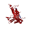
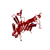
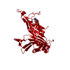
- PDB-1x27: Crystal Structure of Lck SH2-SH3 with SH2 binding site of p130Cas -
+ Open data

- Basic information
| Entry | Database: PDB / ID: 1x27 | ||||||
|---|---|---|---|---|---|---|---|
| Title | Crystal Structure of Lck SH2-SH3 with SH2 binding site of p130Cas | ||||||
 Components Components |
| ||||||
 Keywords Keywords | SIGNALING PROTEIN / Lck-Cas complex / LCK phospho-peptide complex / High affinity LCK-Cas complex | ||||||
| Function / homology |  Function and homology information Function and homology informationp130Cas linkage to MAPK signaling for integrins / PTK6 Regulates RHO GTPases, RAS GTPase and MAP kinases / VEGFA-VEGFR2 Pathway / Downstream signal transduction / antigen receptor-mediated signaling pathway / cellular response to endothelin / regulation of lymphocyte activation / positive regulation of leukocyte cell-cell adhesion / negative regulation of substrate adhesion-dependent cell spreading / CD27 signaling pathway ...p130Cas linkage to MAPK signaling for integrins / PTK6 Regulates RHO GTPases, RAS GTPase and MAP kinases / VEGFA-VEGFR2 Pathway / Downstream signal transduction / antigen receptor-mediated signaling pathway / cellular response to endothelin / regulation of lymphocyte activation / positive regulation of leukocyte cell-cell adhesion / negative regulation of substrate adhesion-dependent cell spreading / CD27 signaling pathway / endothelin receptor signaling pathway / regulation of regulatory T cell differentiation / gamma-delta T cell differentiation / positive regulation of gamma-delta T cell differentiation / Fc-gamma receptor signaling pathway / FLT3 signaling through SRC family kinases / protein antigen binding / positive regulation of T cell receptor signaling pathway / Nef Mediated CD4 Down-regulation / CD4 receptor binding / Nef and signal transduction / hepatocyte growth factor receptor signaling pathway / Co-stimulation by CD28 / Interleukin-2 signaling / positive regulation of heterotypic cell-cell adhesion / CD28 dependent Vav1 pathway / Regulation of KIT signaling / leukocyte migration / cellular response to hepatocyte growth factor stimulus / phospholipase activator activity / Co-inhibition by CTLA4 / Translocation of ZAP-70 to Immunological synapse / Phosphorylation of CD3 and TCR zeta chains / neurotrophin TRK receptor signaling pathway / platelet-derived growth factor receptor signaling pathway / PECAM1 interactions / protein serine/threonine phosphatase activity / CD8 receptor binding / pericentriolar material / hemopoiesis / Generation of second messenger molecules / T cell differentiation / immunological synapse / Co-inhibition by PD-1 / RHOH GTPase cycle / CD28 dependent PI3K/Akt signaling / vascular endothelial growth factor receptor signaling pathway / phosphatidylinositol 3-kinase binding / GPVI-mediated activation cascade / T cell receptor binding / phospholipase binding / T cell costimulation / ruffle / positive regulation of intrinsic apoptotic signaling pathway / phosphotyrosine residue binding / release of sequestered calcium ion into cytosol / cellular response to nitric oxide / positive regulation of endothelial cell migration / SH2 domain binding / cell surface receptor protein tyrosine kinase signaling pathway / Signaling by phosphorylated juxtamembrane, extracellular and kinase domain KIT mutants / T cell activation / integrin-mediated signaling pathway / actin filament organization / B cell receptor signaling pathway / cell chemotaxis / non-specific protein-tyrosine kinase / non-membrane spanning protein tyrosine kinase activity / Signaling by SCF-KIT / platelet activation / SH3 domain binding / positive regulation of T cell activation / epidermal growth factor receptor signaling pathway / Constitutive Signaling by Aberrant PI3K in Cancer / cell-cell junction / insulin receptor signaling pathway / DAP12 signaling / PIP3 activates AKT signaling / Downstream TCR signaling / cell migration / T cell receptor signaling pathway / lamellipodium / actin cytoskeleton / ATPase binding / PI5P, PP2A and IER3 Regulate PI3K/AKT Signaling / actin cytoskeleton organization / protein tyrosine kinase activity / protein phosphatase binding / cell adhesion / intracellular signal transduction / positive regulation of cell migration / membrane raft / response to xenobiotic stimulus / G protein-coupled receptor signaling pathway / signaling receptor binding / protein domain specific binding / axon / focal adhesion / positive regulation of gene expression / protein kinase binding Similarity search - Function | ||||||
| Biological species |  Homo sapiens (human) Homo sapiens (human) | ||||||
| Method |  X-RAY DIFFRACTION / SYNCHROTRON / MOLECULAR REPLACEMENT / Resolution: 2.7 Å X-RAY DIFFRACTION / SYNCHROTRON / MOLECULAR REPLACEMENT / Resolution: 2.7 Å | ||||||
 Authors Authors | Nasertorabi, F. / Tars, K. / Becherer, K. / Kodandapani, R. / Liljas, L. / Vuori, K. / Ely, K.R. | ||||||
 Citation Citation | Journal: J.MOL.RECOG. / Year: 2006 Title: Molecular basis for regulation of Src by the docking protein p130Cas Authors: Nasertorabi, F. / Tars, K. / Becherer, K. / Kodandapani, R. / Liljas, L. / Vuori, K. / Ely, K.R. | ||||||
| History |
|
- Structure visualization
| Structure viewer | Molecule: MolmilJmol/JSmol |
|---|
- Downloads & links
-Download
| PDBx/mmCIF format | 1x27.cif.gz | 209.9 KB | Display | PDBx/mmCIF format |
|---|---|---|---|---|
| PDB format | pdb1x27.ent.gz | 171.5 KB | Display | PDB format |
| PDBx/mmJSON format | 1x27.json.gz | Tree view | PDBx/mmJSON format | |
| Others |  Other downloads Other downloads |
-Validation report
| Arichive directory | https://data.pdbj.org/pub/pdb/validation_reports/x2/1x27ftp://data.pdbj.org/pub/pdb/validation_reports/x2/1x27 | HTTPS FTP |
|---|
-Related structure data
| Related structure data |  1lckS S: Starting model for refinement |
|---|---|
| Similar structure data |
-Links
 PDBj
PDBj


- Assembly
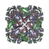
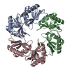
| Deposited unit | 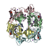
| ||||||||
|---|---|---|---|---|---|---|---|---|---|
| 1 |
| ||||||||
| Unit cell |
|
-Components
| #1: Protein | Mass: 18878.982 Da / Num. of mol.: 6 / Fragment: SH2-SH3 domain Source method: isolated from a genetically manipulated source Source: (gene. exp.) Homo sapiens (human) / Gene: LCK / Plasmid: pET 28a / Species (production host): Escherichia coli / Production host:  #2: Protein/peptide | Mass: 1265.262 Da / Num. of mol.: 6 / Fragment: residues 759-767 / Source method: obtained synthetically Details: The peptide was chemically synthesized. Identical to naturally accured sequence in p130Cas References: UniProt: Q63767 #3: Chemical | ChemComp-NA /   Mass: 22.990 Da / Num. of mol.: 6 / Source method: obtained synthetically / Formula: Na Mass: 22.990 Da / Num. of mol.: 6 / Source method: obtained synthetically / Formula: Na#4: Water | ChemComp-HOH / |  Mass: 18.015 Da / Num. of mol.: 47 / Source method: isolated from a natural source / Formula: H2O Mass: 18.015 Da / Num. of mol.: 47 / Source method: isolated from a natural source / Formula: H2OHas protein modification | Y | |
|---|
-Experimental details
-Experiment
| Experiment | Method: X-RAY DIFFRACTION / Number of used crystals: 1 |
|---|
- Sample preparation
| Crystal | Density Matthews: 2.95 Å3/Da / Density % sol: 58.3 % |
|---|---|
| Crystal grow | Temperature: 295 K / Method: vapor diffusion, hanging drop / pH: 9 Details: N-tris[hydroxymethyl]methyl-3-aminopropane-sulfonic acid, di_potasium hydrogen phosphate, pH 9.0, VAPOR DIFFUSION, HANGING DROP, temperature 295K |
-Data collection
| Diffraction | Mean temperature: 100 K |
|---|---|
| Diffraction source | Source: SYNCHROTRON / Site: ESRF  / Beamline: ID14-1 / Wavelength: 0.934 Å / Beamline: ID14-1 / Wavelength: 0.934 Å |
| Detector | Type: MARRESEARCH / Detector: CCD / Date: Feb 28, 2004 |
| Radiation | Protocol: SINGLE WAVELENGTH / Monochromatic (M) / Laue (L): M / Scattering type: x-ray |
| Radiation wavelength | Wavelength: 0.934 Å / Relative weight: 1 |
| Reflection | Resolution: 2.7→30 Å / Num. all: 37482 / Num. obs: 37482 / % possible obs: 96.6 % / Redundancy: 3.5 % / Biso Wilson estimate: 79 Å2 / Rmerge(I) obs: 0.073 / Net I/σ(I): 7.2 |
| Reflection shell | Resolution: 2.7→2.85 Å / Redundancy: 3.5 % / Mean I/σ(I) obs: 1.5 / Num. unique all: 5316 / Rsym value: 0.482 / % possible all: 96 |
- Processing
| Software |
| ||||||||||||||||||||
|---|---|---|---|---|---|---|---|---|---|---|---|---|---|---|---|---|---|---|---|---|---|
| Refinement | Method to determine structure: MOLECULAR REPLACEMENT Starting model: 1LCK Resolution: 2.7→30 Å / Isotropic thermal model: isotropic / Cross valid method: THROUGHOUT / σ(F): 0 / Stereochemistry target values: Engh & Huber
| ||||||||||||||||||||
| Displacement parameters | Biso mean: 60.3 Å2 | ||||||||||||||||||||
| Refine analyze | Luzzati coordinate error obs: 0.39 Å / Luzzati d res low obs: 5 Å / Luzzati sigma a obs: 0.4 Å | ||||||||||||||||||||
| Refinement step | Cycle: LAST / Resolution: 2.7→30 Å
| ||||||||||||||||||||
| Refine LS restraints |
| ||||||||||||||||||||
| LS refinement shell | Resolution: 2.7→2.87 Å / Rfactor Rfree error: 0.022
|
