 Movie
Movie Controller

[English] 日本語
 Yorodumi
- PDB-3cku: Urate oxidase from aspergillus flavus complexed with its inhibito... -
+ Open data

- Basic information
| Entry | Database: PDB / ID: 3cku | ||||||
|---|---|---|---|---|---|---|---|
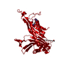
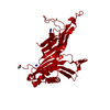
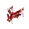
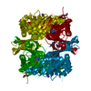
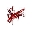
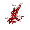
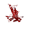
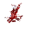
| Title | Urate oxidase from aspergillus flavus complexed with its inhibitor 8-azaxanthin and chloride | ||||||
 Components Components | Uricase | ||||||
 Keywords Keywords | OXIDOREDUCTASE / URIC ACID DEGRADATION / 8-AZAXHANTIN | ||||||
| Function / homology |  Function and homology information Function and homology informationfactor-independent urate hydroxylase / urate oxidase activity / purine nucleobase catabolic process / urate catabolic process / peroxisome Similarity search - Function | ||||||
| Biological species |  | ||||||
| Method |  X-RAY DIFFRACTION / SYNCHROTRON / FOURIER SYNTHESIS / Resolution: 1.7 Å X-RAY DIFFRACTION / SYNCHROTRON / FOURIER SYNTHESIS / Resolution: 1.7 Å | ||||||
 Authors Authors | Colloc'h, N. / Prange, T. | ||||||
 Citation Citation | Journal: Biophys.J. / Year: 2008 Title: Oxygen pressurized X-ray crystallography: probing the dioxygen binding site in cofactorless urate oxidase and implications for its catalytic mechanism. Authors: Colloc'h, N. / Gabison, L. / Monard, G. / Altarsha, M. / Chiadmi, M. / Marassio, G. / Sopkova-de Oliveira Santos, J. / El Hajji, M. / Castro, B. / Abraini, J.H. / Prange, T. #1: Journal: Nat.Struct.Biol. / Year: 1997 Title: Crystal structure of the protein drug urate oxidase-inhibitor complex at 2.05 A resolution. Authors: Colloc'h, N. / el Hajji, M. / Bachet, B. / L'Hermite, G. / Schiltz, M. / Prange, T. / Castro, B. / Mornon, J.P. #2: Journal: Acta Crystallogr.,Sect.D / Year: 2004Title: Complexed and ligand-free high-resolution structures of urate oxidase (Uox) from Aspergillus flavus: a reassignment of the active-site binding mode. Authors: Retailleau, P. / Colloc'h, N. / Vivares, D. / Bonnete, F. / Castro, B. / El-Hajji, M. / Mornon, J.P. / Monard, G. / Prange, T. #3: Journal: Biophys.J. / Year: 2007Title: Protein crystallography under xenon and nitrous oxide pressure: comparison with in vivo pharmacology studies and implications for the mechanism of inhaled anesthetic action. Authors: Colloc'h, N. / Sopkova-de Oliveira Santos, J. / Retailleau, P. / Vivares, D. / Bonnete, F. / Langlois d'Estainto, B. / Gallois, B. / Brisson, A. / Risso, J.J. / Lemaire, M. / Prange, T. / Abraini, J.H. | ||||||
| History |
|
- Structure visualization
| Structure viewer | Molecule: MolmilJmol/JSmol |
|---|
- Downloads & links
-Download
| PDBx/mmCIF format | 3cku.cif.gz | 80.6 KB | Display | PDBx/mmCIF format |
|---|---|---|---|---|
| PDB format | pdb3cku.ent.gz | 59.1 KB | Display | PDB format |
| PDBx/mmJSON format | 3cku.json.gz | Tree view | PDBx/mmJSON format | |
| Others |  Other downloads Other downloads |
-Validation report
| Arichive directory | https://data.pdbj.org/pub/pdb/validation_reports/ck/3ckuftp://data.pdbj.org/pub/pdb/validation_reports/ck/3cku | HTTPS FTP |
|---|
-Related structure data
| Related structure data |  2zkaC  2zkbC  3cksC 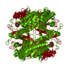 1r4sS S: Starting model for refinement C: citing same article ( |
|---|---|
| Similar structure data |


-Links
 PDBj
PDBj
- Assembly
| Deposited unit | 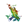
| ||||||||
|---|---|---|---|---|---|---|---|---|---|
| 1 | 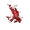
| ||||||||
| Unit cell |
|
-Components
| #1: Protein | Mass: 34183.590 Da / Num. of mol.: 1 Source method: isolated from a genetically manipulated source Source: (gene. exp.)  References: UniProt: Q00511, factor-independent urate hydroxylase |
|---|---|
| #2: Chemical | ChemComp-CL /   Mass: 35.453 Da / Num. of mol.: 1 / Source method: obtained synthetically / Formula: Cl Mass: 35.453 Da / Num. of mol.: 1 / Source method: obtained synthetically / Formula: Cl |
| #3: Chemical | ChemComp-NA /   Mass: 22.990 Da / Num. of mol.: 1 / Source method: obtained synthetically / Formula: Na Mass: 22.990 Da / Num. of mol.: 1 / Source method: obtained synthetically / Formula: Na |
| #4: Chemical | ChemComp-AZA /   Mass: 153.099 Da / Num. of mol.: 1 / Source method: obtained synthetically / Formula: C4H3N5O2 Mass: 153.099 Da / Num. of mol.: 1 / Source method: obtained synthetically / Formula: C4H3N5O2 |
| #5: Water | ChemComp-HOH /  Mass: 18.015 Da / Num. of mol.: 275 / Source method: isolated from a natural source / Formula: H2O Mass: 18.015 Da / Num. of mol.: 275 / Source method: isolated from a natural source / Formula: H2O |
| Has protein modification | Y |
-Experimental details
-Experiment
| Experiment | Method: X-RAY DIFFRACTION / Number of used crystals: 1 |
|---|
- Sample preparation
| Crystal | Density Matthews: 2.87 Å3/Da / Density % sol: 57.08 % |
|---|---|
| Crystal grow | Temperature: 291 K / Method: vapor diffusion, sitting drop / pH: 8 Details: 0.2MG/ML 8-AZAXANTHIN, 0.3M SODIUM CHLORIDE, 0.05M TRIS/HCL, 10-15% W/V PEG 8000, VAPOR DIFFUSION, SITTING DROP, temperature 291K |
-Data collection
| Diffraction | Mean temperature: 100 K |
|---|---|
| Diffraction source | Source: SYNCHROTRON / Site: ESRF  / Beamline: BM14 / Wavelength: 0.964 / Wavelength: 0.964 Å / Beamline: BM14 / Wavelength: 0.964 / Wavelength: 0.964 Å |
| Detector | Type: MARMOSAIC 225 mm CCD / Detector: CCD / Date: Mar 12, 2007 / Details: BENT MIRROR |
| Radiation | Monochromator: SI(111) / Protocol: SINGLE WAVELENGTH / Monochromatic (M) / Laue (L): M / Scattering type: x-ray |
| Radiation wavelength | Wavelength: 0.964 Å / Relative weight: 1 |
| Reflection | Resolution: 1.7→10 Å / Num. all: 43251 / Num. obs: 42048 / % possible obs: 99.63 % / Observed criterion σ(F): 2 / Observed criterion σ(I): 4 / Redundancy: 3.5 % / Biso Wilson estimate: 20.3 Å2 / Rmerge(I) obs: 0.0555 / Rsym value: 0.0531 / Net I/σ(I): 22 |
| Reflection shell | Resolution: 1.7→1.79 Å / Redundancy: 3.5 % / Rmerge(I) obs: 0.099 / Mean I/σ(I) obs: 13.7 / Num. unique all: 6302 / Rsym value: 0.084 / % possible all: 100 |
- Processing
| Software |
| |||||||||||||||||||||||||||||||||
|---|---|---|---|---|---|---|---|---|---|---|---|---|---|---|---|---|---|---|---|---|---|---|---|---|---|---|---|---|---|---|---|---|---|---|
| Refinement | Method to determine structure: FOURIER SYNTHESIS Starting model: 1R4S Resolution: 1.7→10 Å / Num. parameters: 10759 / Num. restraintsaints: 9913 / Cross valid method: THROUGHOUT / σ(F): 2 / σ(I): 4 / Stereochemistry target values: ENGH & HUBER
| |||||||||||||||||||||||||||||||||
| Refine analyze | Num. disordered residues: 10 / Occupancy sum hydrogen: 0 / Occupancy sum non hydrogen: 2651 | |||||||||||||||||||||||||||||||||
| Refinement step | Cycle: LAST / Resolution: 1.7→10 Å
| |||||||||||||||||||||||||||||||||
| Refine LS restraints |
|
