 Movie
Movie Controller

[English] 日本語
 Yorodumi
- PDB-1wyv: Crystal structure of glycine decarboxylase (P-protein) of the gly... -
+ Open data

- Basic information
| Entry | Database: PDB / ID: 1wyv | ||||||
|---|---|---|---|---|---|---|---|
| Title | Crystal structure of glycine decarboxylase (P-protein) of the glycine cleavage system, in inhibitor-bound form | ||||||
 Components Components |
| ||||||
 Keywords Keywords | OXIDOREDUCTASE / ALPHA(2)BETA(2) TETRAMER / RIKEN Structural Genomics/Proteomics Initiative / RSGI / Structural Genomics | ||||||
| Function / homology |  Function and homology information Function and homology informationglycine dehydrogenase (aminomethyl-transferring) / glycine dehydrogenase (decarboxylating) activity / glycine decarboxylation via glycine cleavage system / glycine cleavage complex / nucleoside metabolic process / glycine binding / pyridoxal phosphate binding / cytosol Similarity search - Function | ||||||
| Biological species |   Thermus thermophilus (bacteria) Thermus thermophilus (bacteria) | ||||||
| Method |  X-RAY DIFFRACTION / SYNCHROTRON / FOURIER SYNTHESIS / Resolution: 2.4 Å X-RAY DIFFRACTION / SYNCHROTRON / FOURIER SYNTHESIS / Resolution: 2.4 Å | ||||||
 Authors Authors | Nakai, T. / Nakagawa, N. / Maoka, N. / Masui, R. / Kuramitsu, S. / Kamiya, N. / RIKEN Structural Genomics/Proteomics Initiative (RSGI) | ||||||
 Citation Citation | Journal: Embo J. / Year: 2005 Title: Structure of P-protein of the glycine cleavage system: implications for nonketotic hyperglycinemia Authors: Nakai, T. / Nakagawa, N. / Maoka, N. / Masui, R. / Kuramitsu, S. / Kamiya, N. | ||||||
| History |
|
- Structure visualization
| Structure viewer | Molecule: MolmilJmol/JSmol |
|---|
- Downloads & links
-Download
| PDBx/mmCIF format | 1wyv.cif.gz | 710.7 KB | Display | PDBx/mmCIF format |
|---|---|---|---|---|
| PDB format | pdb1wyv.ent.gz | 580.3 KB | Display | PDB format |
| PDBx/mmJSON format | 1wyv.json.gz | Tree view | PDBx/mmJSON format | |
| Others |  Other downloads Other downloads |
-Validation report
| Arichive directory | https://data.pdbj.org/pub/pdb/validation_reports/wy/1wyvftp://data.pdbj.org/pub/pdb/validation_reports/wy/1wyv | HTTPS FTP |
|---|
-Related structure data


-Links
 PDBj
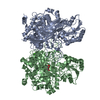
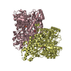
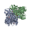
PDBj- Assembly
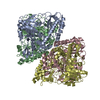
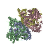
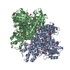
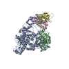
| Deposited unit | 
| ||||||||
|---|---|---|---|---|---|---|---|---|---|
| 1 | 
| ||||||||
| 2 | 
| ||||||||
| Unit cell |
| ||||||||
| Details | The biological assembly is an alpha(2)beta(2) tetramer. The asymmetric unit contains two sets of the assembly (chains A-D and E-H). |
-Components
| #1: Protein | Mass: 47168.133 Da / Num. of mol.: 4 Source method: isolated from a genetically manipulated source Source: (gene. exp.) Thermus thermophilus (bacteria) / Strain: HB8 / Gene: GCSA / Plasmid: pET11A / Production host: References: GenBank: 55771907, UniProt: Q5SKW8*PLUS, glycine dehydrogenase (aminomethyl-transferring) #2: Protein | Mass: 52784.711 Da / Num. of mol.: 4 Source method: isolated from a genetically manipulated source Source: (gene. exp.) Thermus thermophilus (bacteria) / Strain: HB8 / Gene: GCSB / Plasmid: pET11A / Production host: References: GenBank: 55771908, UniProt: Q5SKW7*PLUS, glycine dehydrogenase (aminomethyl-transferring) #3: Chemical | ChemComp-PLP /   Mass: 247.142 Da / Num. of mol.: 4 / Source method: obtained synthetically / Formula: C8H10NO6P Mass: 247.142 Da / Num. of mol.: 4 / Source method: obtained synthetically / Formula: C8H10NO6P#4: Chemical | ChemComp-AOA / ( 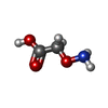  Mass: 91.066 Da / Num. of mol.: 4 / Source method: obtained synthetically / Formula: C2H5NO3 Mass: 91.066 Da / Num. of mol.: 4 / Source method: obtained synthetically / Formula: C2H5NO3#5: Water | ChemComp-HOH / |  Mass: 18.015 Da / Num. of mol.: 1192 / Source method: isolated from a natural source / Formula: H2O Mass: 18.015 Da / Num. of mol.: 1192 / Source method: isolated from a natural source / Formula: H2O |
|---|
-Experimental details
-Experiment
| Experiment | Method: X-RAY DIFFRACTION / Number of used crystals: 1 |
|---|
- Sample preparation
| Crystal | Density Matthews: 2.69 Å3/Da / Density % sol: 54 % |
|---|---|
| Crystal grow | Temperature: 277 K / Method: vapor diffusion, hanging drop / pH: 6.5 Details: PEG 3350, lithium sulfate, MES, pH 6.5, VAPOR DIFFUSION, HANGING DROP, temperature 277K |
-Data collection
| Diffraction | Mean temperature: 90 K |
|---|---|
| Diffraction source | Source: SYNCHROTRON / Site: SPring-8  / Beamline: BL44B2 / Wavelength: 1 Å / Beamline: BL44B2 / Wavelength: 1 Å |
| Detector | Type: ADSC QUANTUM 210 / Detector: CCD / Date: Jun 29, 2004 |
| Radiation | Monochromator: SI(111) / Protocol: SINGLE WAVELENGTH / Monochromatic (M) / Laue (L): M / Scattering type: x-ray |
| Radiation wavelength | Wavelength: 1 Å / Relative weight: 1 |
| Reflection | Resolution: 2.4→50 Å / Num. all: 167358 / Num. obs: 166371 / % possible obs: 99.4 % / Observed criterion σ(I): 0 / Redundancy: 6.6 % / Biso Wilson estimate: 32.7 Å2 / Rmerge(I) obs: 0.118 / Net I/σ(I): 16 |
| Reflection shell | Resolution: 2.4→2.49 Å / Redundancy: 5.7 % / Rmerge(I) obs: 0.578 / Mean I/σ(I) obs: 3.1 / Num. unique all: 16003 / % possible all: 96.6 |
- Processing
| Software |
| ||||||||||||||||||||||||||||||||||||||||||||||||||||||||||||||||||||||||||||||||
|---|---|---|---|---|---|---|---|---|---|---|---|---|---|---|---|---|---|---|---|---|---|---|---|---|---|---|---|---|---|---|---|---|---|---|---|---|---|---|---|---|---|---|---|---|---|---|---|---|---|---|---|---|---|---|---|---|---|---|---|---|---|---|---|---|---|---|---|---|---|---|---|---|---|---|---|---|---|---|---|---|---|
| Refinement | Method to determine structure: FOURIER SYNTHESIS Starting model: P-PROTEIN IN THE HOLO-FORM Resolution: 2.4→47.9 Å / Rfactor Rfree error: 0.003 / Data cutoff high absF: 4234196.18 / Data cutoff low absF: 0 / Isotropic thermal model: RESTRAINED / Cross valid method: THROUGHOUT / σ(F): 0 / Stereochemistry target values: Engh & Huber
| ||||||||||||||||||||||||||||||||||||||||||||||||||||||||||||||||||||||||||||||||
| Solvent computation | Solvent model: FLAT MODEL / Bsol: 36.302 Å2 / ksol: 0.353867 e/Å3 | ||||||||||||||||||||||||||||||||||||||||||||||||||||||||||||||||||||||||||||||||
| Displacement parameters | Biso mean: 35.6 Å2
| ||||||||||||||||||||||||||||||||||||||||||||||||||||||||||||||||||||||||||||||||
| Refine analyze |
| ||||||||||||||||||||||||||||||||||||||||||||||||||||||||||||||||||||||||||||||||
| Refinement step | Cycle: LAST / Resolution: 2.4→47.9 Å
| ||||||||||||||||||||||||||||||||||||||||||||||||||||||||||||||||||||||||||||||||
| Refine LS restraints |
| ||||||||||||||||||||||||||||||||||||||||||||||||||||||||||||||||||||||||||||||||
| LS refinement shell | Resolution: 2.4→2.55 Å / Rfactor Rfree error: 0.008 / Total num. of bins used: 6
| ||||||||||||||||||||||||||||||||||||||||||||||||||||||||||||||||||||||||||||||||
| Xplor file |
|
