- PDB-1wu3: Crystal structure of recombinant murine interferon beta -
+
Open data
ID or keywords:
Loading...
-
Basic information
Entry
Database: PDB / ID: 1wu3
Title
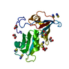
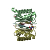
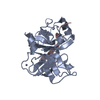
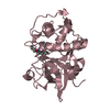
Crystal structure of recombinant murine interferon beta
Components
Interferon beta
Keywords
CYTOKINE / alpha-helix-bundle
Function / homology
Function and homology information
negative regulation of Lewy body formation / negative regulation of mononuclear cell migration / Regulation of IFNA/IFNB signaling / negative regulation of matrix metallopeptidase secretion / type I interferon receptor binding / B cell activation involved in immune response / chloramphenicol O-acetyltransferase activity / negative regulation of immunoglobulin production / negative regulation of T cell differentiation / Interferon alpha/beta signaling ...negative regulation of Lewy body formation / negative regulation of mononuclear cell migration / Regulation of IFNA/IFNB signaling / negative regulation of matrix metallopeptidase secretion / type I interferon receptor binding / B cell activation involved in immune response / chloramphenicol O-acetyltransferase activity / negative regulation of immunoglobulin production / negative regulation of T cell differentiation / Interferon alpha/beta signaling / natural killer cell activation involved in immune response / negative regulation of blood-brain barrier permeability / negative regulation of neuroinflammatory response / positive regulation of transforming growth factor beta production / negative regulation of T-helper 2 cell cytokine production / negative regulation of cell adhesion molecule production / T cell activation involved in immune response / cytokine receptor binding / macrophage activation involved in immune response / cell surface receptor signaling pathway via STAT / cellular response to dsRNA / negative regulation of viral genome replication / type I interferon-mediated signaling pathway / response to exogenous dsRNA / negative regulation of osteoclast differentiation / negative regulation of type II interferon production / humoral immune response / B cell proliferation / cellular response to dexamethasone stimulus / cell surface receptor signaling pathway via JAK-STAT / cellular response to interferon-beta / positive regulation of autophagy / antiviral innate immune response / cytokine activity / positive regulation of apoptotic signaling pathway / neuron cellular homeostasis / cellular response to virus / positive regulation of protein localization to nucleus / cytokine-mediated signaling pathway / defense response to virus / adaptive immune response / defense response to bacterium / negative regulation of cell population proliferation / innate immune response / regulation of transcription by RNA polymerase II / positive regulation of transcription by RNA polymerase II / : / extracellular region Similarity search - Function
#3: Journal: Pharmacol.Ther. / Year: 1993 Title: Structural, functional and evolutionary implications of the three-dimensional crystal structure of murine interferon-beta Authors: Mitsui, Y. / Senda, T. / Shimazu, T. / Matsuda, S. / Utsumi, J.
Method to determine structure: MIR / Resolution: 2.15→7.95 Å / Cor.coef. Fo:Fc: 0.953 / Cor.coef. Fo:Fc free: 0.915 / SU B: 6.918 / SU ML: 0.169 / Cross valid method: THROUGHOUT / σ(F): 0 / ESU R: 0.233 / ESU R Free: 0.203 / Stereochemistry target values: MAXIMUM LIKELIHOOD / Details: HYDROGENS HAVE BEEN ADDED IN THE RIDING POSITIONS
Rfactor
Num. reflection
% reflection
Selection details
Rfree
0.24731
1182
10.1 %
RANDOM
Rwork
0.19162
-
-
-
all
0.1973
10525
-
-
obs
0.1973
10525
100 %
-
Solvent computation
Ion probe radii: 0.8 Å / Shrinkage radii: 0.8 Å / VDW probe radii: 1.2 Å / Solvent model: BABINET MODEL WITH MASK
Displacement parameters
Biso mean: 38.011 Å2
Baniso -1
Baniso -2
Baniso -3
1-
0.25 Å2
0.12 Å2
0 Å2
2-
-
0.25 Å2
0 Å2
3-
-
-
-0.37 Å2
Refinement step
Cycle: LAST / Resolution: 2.15→7.95 Å
Protein
Nucleic acid
Ligand
Solvent
Total
Num. atoms
1394
0
0
48
1442
Refine LS restraints
Refine-ID
Type
Dev ideal
Dev ideal target
Number
X-RAY DIFFRACTION
r_bond_refined_d
0.012
0.022
1412
X-RAY DIFFRACTION
r_angle_refined_deg
1.375
1.94
1899
X-RAY DIFFRACTION
r_dihedral_angle_1_deg
3.181
5
159
X-RAY DIFFRACTION
r_dihedral_angle_2_deg
39.763
23.553
76
X-RAY DIFFRACTION
r_dihedral_angle_3_deg
19.054
15
283
X-RAY DIFFRACTION
r_dihedral_angle_4_deg
26.3
15
13
X-RAY DIFFRACTION
r_chiral_restr
0.093
0.2
208
X-RAY DIFFRACTION
r_gen_planes_refined
0.006
0.02
1044
X-RAY DIFFRACTION
r_nbd_refined
0.227
0.2
671
X-RAY DIFFRACTION
r_nbtor_refined
0.316
0.2
995
X-RAY DIFFRACTION
r_xyhbond_nbd_refined
0.108
0.2
45
X-RAY DIFFRACTION
r_symmetry_vdw_refined
0.217
0.2
35
X-RAY DIFFRACTION
r_symmetry_hbond_refined
0.081
0.2
1
X-RAY DIFFRACTION
r_mcbond_it
1.848
1.5
819
X-RAY DIFFRACTION
r_mcangle_it
2.425
2
1300
X-RAY DIFFRACTION
r_scbond_it
3.145
3
680
X-RAY DIFFRACTION
r_scangle_it
4.84
4.5
599
LS refinement shell
Resolution: 2.15→2.202 Å / Total num. of bins used: 20
Rfactor
Num. reflection
% reflection
Rfree
0.361
81
-
Rwork
0.345
604
-
obs
-
-
100 %
+
About Yorodumi
-
News
-
Feb 9, 2022. New format data for meta-information of EMDB entries
New format data for meta-information of EMDB entries
Version 3 of the EMDB header file is now the official format.
The previous official version 1.9 will be removed from the archive.
In the structure databanks used in Yorodumi, some data are registered as the other names, "COVID-19 virus" and "2019-nCoV". Here are the details of the virus and the list of structure data.
Jan 31, 2019. EMDB accession codes are about to change! (news from PDBe EMDB page)
EMDB accession codes are about to change! (news from PDBe EMDB page)
The allocation of 4 digits for EMDB accession codes will soon come to an end. Whilst these codes will remain in use, new EMDB accession codes will include an additional digit and will expand incrementally as the available range of codes is exhausted. The current 4-digit format prefixed with “EMD-” (i.e. EMD-XXXX) will advance to a 5-digit format (i.e. EMD-XXXXX), and so on. It is currently estimated that the 4-digit codes will be depleted around Spring 2019, at which point the 5-digit format will come into force.
The EM Navigator/Yorodumi systems omit the EMD- prefix.
Related info.:Q: What is EMD? / ID/Accession-code notation in Yorodumi/EM Navigator
Yorodumi is a browser for structure data from EMDB, PDB, SASBDB, etc.
This page is also the successor to EM Navigator detail page, and also detail information page/front-end page for Omokage search.
The word "yorodu" (or yorozu) is an old Japanese word meaning "ten thousand". "mi" (miru) is to see.
Related info.:EMDB / PDB / SASBDB / Comparison of 3 databanks / Yorodumi Search / Aug 31, 2016. New EM Navigator & Yorodumi / Yorodumi Papers / Jmol/JSmol / Function and homology information / Changes in new EM Navigator and Yorodumi
 Movie
Movie Controller


 Open data
 Basic information
 Components
 Keywords
 Function and homology information
 X-RAY DIFFRACTION /
  Authors
 Citation
 Structure visualization
 Downloads & links
 Other downloads
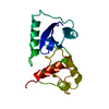


 PDBj
PDBj


 Assembly
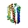

 Mass: 18.015 Da / Num. of mol.: 48 / Source method: isolated from a natural source / Formula: H2O
 Sample preparation
 / Beamline: BL-6A / Wavelength: 1.04 Å
 Processing