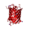
 Movie
Movie Controller

[English] 日本語
 Yorodumi
- PDB-1wo8: Crystal structure of methylglyoxal synthase from Thermus thermoph... -
+ Open data

- Basic information
| Entry | Database: PDB / ID: 1wo8 | ||||||
|---|---|---|---|---|---|---|---|
| Title | Crystal structure of methylglyoxal synthase from Thermus thermophilus HB8 | ||||||
 Components Components | methylglyoxal synthase | ||||||
 Keywords Keywords | LYASE / methylglyoxal synthase / Thermus thermophilus HB8 / Structural Genomics / RIKEN Structural Genomics/Proteomics Initiative / RSGI | ||||||
| Function / homology |  Function and homology information Function and homology informationmethylglyoxal biosynthetic process / methylglyoxal synthase / methylglyoxal synthase activity / cytosol Similarity search - Function | ||||||
| Biological species |   Thermus thermophilus HB8 (bacteria) Thermus thermophilus HB8 (bacteria) | ||||||
| Method |  X-RAY DIFFRACTION / SYNCHROTRON / SAD / Resolution: 1.7 Å X-RAY DIFFRACTION / SYNCHROTRON / SAD / Resolution: 1.7 Å | ||||||
 Authors Authors | Sugahara, M. / Kunishima, N. / RIKEN Structural Genomics/Proteomics Initiative (RSGI) | ||||||
 Citation Citation | Journal: To be Published Title: Crystal structure of methylglyoxal synthase from Thermus thermophilus HB8 Authors: Sugahara, M. / Kunishima, N. | ||||||
| History |
|
- Structure visualization
| Structure viewer | Molecule: MolmilJmol/JSmol |
|---|
- Downloads & links
-Download
| PDBx/mmCIF format | 1wo8.cif.gz | 163.5 KB | Display | PDBx/mmCIF format |
|---|---|---|---|---|
| PDB format | pdb1wo8.ent.gz | 130.1 KB | Display | PDB format |
| PDBx/mmJSON format | 1wo8.json.gz | Tree view | PDBx/mmJSON format | |
| Others |  Other downloads Other downloads |
-Validation report
| Arichive directory | https://data.pdbj.org/pub/pdb/validation_reports/wo/1wo8ftp://data.pdbj.org/pub/pdb/validation_reports/wo/1wo8 | HTTPS FTP |
|---|
-Related structure data
| Similar structure data | |
|---|---|
| Other databases |

-Links
 PDBj
PDBj
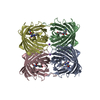
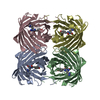
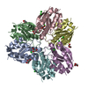
- Assembly
| Deposited unit | 
| ||||||||||||
|---|---|---|---|---|---|---|---|---|---|---|---|---|---|
| 1 |
| ||||||||||||
| 2 | 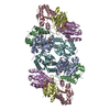
| ||||||||||||
| Unit cell |
| ||||||||||||
| Components on special symmetry positions |
| ||||||||||||
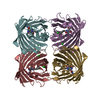
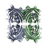
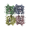
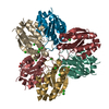
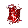
| Details | The biological assembly is a hexamer(chain A,B,C,D,E,F) in the asymmetric unit. |
-Components
| #1: Protein | Mass: 13397.733 Da / Num. of mol.: 6 Source method: isolated from a genetically manipulated source Source: (gene. exp.) Thermus thermophilus HB8 (bacteria) / Species: Thermus thermophilus / Strain: HB8 / ATCC 27634 / DSM 579 / Plasmid: pET11a / Production host: #2: Chemical | ChemComp-SO4 /   Mass: 96.063 Da / Num. of mol.: 7 / Source method: obtained synthetically / Formula: SO4 Mass: 96.063 Da / Num. of mol.: 7 / Source method: obtained synthetically / Formula: SO4#3: Water | ChemComp-HOH / |  Mass: 18.015 Da / Num. of mol.: 816 / Source method: isolated from a natural source / Formula: H2O Mass: 18.015 Da / Num. of mol.: 816 / Source method: isolated from a natural source / Formula: H2O |
|---|
-Experimental details
-Experiment
| Experiment | Method: X-RAY DIFFRACTION / Number of used crystals: 2 |
|---|
- Sample preparation
| Crystal | Density Matthews: 3 Å3/Da / Density % sol: 58.2 % |
|---|---|
| Crystal grow | Temperature: 291 K / Method: microbatch / pH: 4.6 Details: Lithium sulfate, pH 4.6, microbatch, temperature 291K |
-Data collection
| Diffraction |
| ||||||||||||||||||
|---|---|---|---|---|---|---|---|---|---|---|---|---|---|---|---|---|---|---|---|
| Diffraction source |
| ||||||||||||||||||
| Detector |
| ||||||||||||||||||
| Radiation | Protocol: SINGLE WAVELENGTH / Monochromatic (M) / Laue (L): M / Scattering type: x-ray | ||||||||||||||||||
| Radiation wavelength |
| ||||||||||||||||||
| Reflection | Resolution: 1.7→40 Å / Num. all: 104491 / Num. obs: 104491 / % possible obs: 99.9 % / Observed criterion σ(F): 0 / Observed criterion σ(I): 0 / Redundancy: 8.9 % / Biso Wilson estimate: 19.44 Å2 / Rmerge(I) obs: 0.106 / Rsym value: 0.1 / Net I/σ(I): 6.8 | ||||||||||||||||||
| Reflection shell | Resolution: 1.7→1.76 Å / Redundancy: 8.5 % / Rmerge(I) obs: 0.432 / Mean I/σ(I) obs: 3.1 / Num. unique all: 10286 / Rsym value: 0.405 / % possible all: 99.4 |

- Processing
| Software |
| |||||||||||||||||||||||||
|---|---|---|---|---|---|---|---|---|---|---|---|---|---|---|---|---|---|---|---|---|---|---|---|---|---|---|
| Refinement | Method to determine structure: SAD / Resolution: 1.7→39.1 Å / Isotropic thermal model: anisotropic / Cross valid method: THROUGHOUT / σ(F): 0 / Stereochemistry target values: Engh & Huber
| |||||||||||||||||||||||||
| Displacement parameters | Biso mean: 21.7 Å2
| |||||||||||||||||||||||||
| Refine analyze |
| |||||||||||||||||||||||||
| Refinement step | Cycle: LAST / Resolution: 1.7→39.1 Å
| |||||||||||||||||||||||||
| Refine LS restraints |
| |||||||||||||||||||||||||
| LS refinement shell | Resolution: 1.7→1.76 Å / Rfactor Rfree error: 0.012
|
