 Movie
Movie Controller

[English] 日本語
 Yorodumi
- PDB-1wgu: Solution Structure of the C-terminal Phosphotyrosine Interaction ... -
+ Open data

- Basic information
| Entry | Database: PDB / ID: 1wgu | ||||||
|---|---|---|---|---|---|---|---|
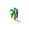
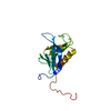
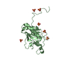
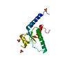
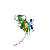
| Title | Solution Structure of the C-terminal Phosphotyrosine Interaction Domain of APBB2 from Mouse | ||||||
 Components Components | amyloid beta (A4) precursor protein-binding, family B, member 2 | ||||||
 Keywords Keywords | PROTEIN BINDING / phosphotyrosine-interaction domain / amyloid disease / structural genomics / RIKEN Structural Genomics/Proteomics Initiative / RSGI | ||||||
| Function / homology |  Function and homology information Function and homology informationnegative regulation of cell cycle phase transition / maintenance of lens transparency / smooth muscle contraction / extracellular matrix organization / axon guidance / neuron migration / amyloid-beta binding / early endosome / protein stabilization / positive regulation of apoptotic process ...negative regulation of cell cycle phase transition / maintenance of lens transparency / smooth muscle contraction / extracellular matrix organization / axon guidance / neuron migration / amyloid-beta binding / early endosome / protein stabilization / positive regulation of apoptotic process / negative regulation of apoptotic process / endoplasmic reticulum / negative regulation of transcription by RNA polymerase II / Golgi apparatus / positive regulation of transcription by RNA polymerase II / nucleus / cytoplasm Similarity search - Function | ||||||
| Biological species |  | ||||||
| Method | SOLUTION NMR / torsion angle dynamics | ||||||
 Authors Authors | Li, H. / Hayashi, F. / Koshiba, S. / Inoue, M. / Kigawa, T. / Yokoyama, S. / RIKEN Structural Genomics/Proteomics Initiative (RSGI) | ||||||
 Citation Citation | Journal: J.Biol.Chem. / Year: 2008 Title: Structure of the C-terminal phosphotyrosine interaction domain of Fe65L1 complexed with the cytoplasmic tail of amyloid precursor protein reveals a novel peptide binding mode Authors: Li, H. / Koshiba, S. / Hayashi, F. / Tochio, N. / Tomizawa, T. / Kasai, T. / Yabuki, T. / Motoda, Y. / Harada, T. / Watanabe, S. / Inoue, M. / Hayashizaki, Y. / Tanaka, A. / Kigawa, T. / Yokoyama, S. | ||||||
| History |
| ||||||
| Remark 650 | HELIX DETERMINATION METHOD: AUTHOR DETERMINED | ||||||
| Remark 700 | SHEET DETERMINATION METHOD: AUTHOR DETERMINED |
- Structure visualization
| Structure viewer | Molecule: MolmilJmol/JSmol |
|---|
- Downloads & links
-Download
| PDBx/mmCIF format | 1wgu.cif.gz | 781 KB | Display | PDBx/mmCIF format |
|---|---|---|---|---|
| PDB format | pdb1wgu.ent.gz | 655.6 KB | Display | PDB format |
| PDBx/mmJSON format | 1wgu.json.gz | Tree view | PDBx/mmJSON format | |
| Others |  Other downloads Other downloads |
-Validation report
| Summary document | 1wgu_validation.pdf.gz | 344.8 KB | Display | wwPDB validaton report |
|---|---|---|---|---|
| Full document | 1wgu_full_validation.pdf.gz | 498.8 KB | Display | |
| Data in XML | 1wgu_validation.xml.gz | 59.7 KB | Display | |
| Data in CIF | 1wgu_validation.cif.gz | 73.8 KB | Display | |
| Arichive directory | https://data.pdbj.org/pub/pdb/validation_reports/wg/1wguftp://data.pdbj.org/pub/pdb/validation_reports/wg/1wgu | HTTPS FTP |
-Related structure data
| Related structure data |  2rozC  2yszC  2yt0C 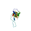 2yt1C C: citing same article ( |
|---|---|
| Similar structure data | |
| Other databases |


-Links
 PDBj
PDBj

- Assembly
| Deposited unit | 
| |||||||||
|---|---|---|---|---|---|---|---|---|---|---|
| 1 |
| |||||||||
| NMR ensembles |
|
-Components
| #1: Protein | Mass: 14750.546 Da / Num. of mol.: 1 / Fragment: Phosphotyrosine-Interaction Domain Source method: isolated from a genetically manipulated source Source: (gene. exp.) |
|---|
-Experimental details
-Experiment
| Experiment | Method: SOLUTION NMR | ||||||||||||
|---|---|---|---|---|---|---|---|---|---|---|---|---|---|
| NMR experiment |
|
- Sample preparation
| Details | Contents: 0.84mM phosphotyrosine-interaction domain U-13C, 15N; 20mM d-Tris-HCl (PH7.0); 100mM NaCl; 2mM d-DTT; 0.02% NaN3; 10% D2O Solvent system: 90% H2O/10% D2O |
|---|---|
| Sample conditions | Ionic strength: 120mM / pH: 7 / Pressure: ambient / Temperature: 298 K |
-NMR measurement
| Radiation | Protocol: SINGLE WAVELENGTH / Monochromatic (M) / Laue (L): M |
|---|---|
| Radiation wavelength | Relative weight: 1 |
| NMR spectrometer | Type: Varian INOVA / Manufacturer: Varian / Model: INOVA / Field strength: 900 MHz |
- Processing
| NMR software |
| ||||||||||||||||||||||||||||
|---|---|---|---|---|---|---|---|---|---|---|---|---|---|---|---|---|---|---|---|---|---|---|---|---|---|---|---|---|---|
| Refinement | Method: torsion angle dynamics / Software ordinal: 1 | ||||||||||||||||||||||||||||
| NMR representative | Selection criteria: lowest energy | ||||||||||||||||||||||||||||
| NMR ensemble | Conformer selection criteria: structures with the least restraint violations, target function Conformers calculated total number: 100 / Conformers submitted total number: 20 |
 NMRPipe