- PDB-1w8i: The Structure of gene product af1683 from Archaeoglobus fulgidus. -
+
Open data
ID or keywords:
Loading...
-
Basic information
Entry
Database: PDB / ID: 1w8i
Title
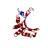
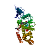
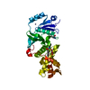
The Structure of gene product af1683 from Archaeoglobus fulgidus.
Components
PUTATIVE VAPC RIBONUCLEASE AF_1683
Keywords
STRUCTURAL GENOMICS / UNKNOWN FUNCTION / ARCHAEOGLOBUS FULGIDUS / HYPOTHETICAL PROTEIN / PSI / PROTEIN STRUCTURE INITIATIVE / MCSG / MIDWEST CENTER FOR STRUCTURAL GENOMICS
Function / homology
Function and homology information
rRNA catabolic process / RNA endonuclease activity / Hydrolases; Acting on ester bonds / hydrolase activity / magnesium ion binding Similarity search - Function
Resolution: 2.1→50 Å / Num. obs: 26396 / % possible obs: 95.8 % / Observed criterion σ(I): 0 / Redundancy: 7.6 % / Rmerge(I) obs: 0.07 / Net I/σ(I): 9.8
Reflection shell
Resolution: 2.1→2.18 Å / Redundancy: 7.6 % / Rmerge(I) obs: 0.6 / Mean I/σ(I) obs: 3.5 / % possible all: 100
-
Processing
Software
Name
Version
Classification
DM
modelbuilding
HKL-2000
datareduction
HKL-3000
phasing
SHELXD
phasing
MLPHARE
phasing
DM
phasing
RESOLVE
phasing
ARP/wARP
phasing
REFMAC
5.2.0005
refinement
Refinement
Method to determine structure: MAD / Resolution: 2.1→57.83 Å / Cor.coef. Fo:Fc: 0.941 / Cor.coef. Fo:Fc free: 0.936 / SU B: 4.013 / SU ML: 0.108 / Cross valid method: THROUGHOUT / ESU R: 0.185 / ESU R Free: 0.152 Stereochemistry target values: MAXIMUM LIKELIHOOD WITH PHASES Details: HYDROGENS HAVE BEEN ADDED IN THE RIDING POSITIONS
Rfactor
Num. reflection
% reflection
Selection details
Rfree
0.225
742
5 %
RANDOM
Rwork
0.211
-
-
-
obs
0.212
13960
99.8 %
-
Solvent computation
Ion probe radii: 0.8 Å / Shrinkage radii: 0.8 Å / VDW probe radii: 1.2 Å / Solvent model: MASK
 Movie
Movie Controller

 Yorodumi
 Open data
 Basic information
 Components
 Keywords
 Function and homology information
 ARCHAEOGLOBUS FULGIDUS (archaea)
 X-RAY DIFFRACTION /
  Authors
 Citation
 Structure visualization
 Downloads & links
 Other downloads


 PDBj
PDBj

 Assembly
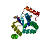


 Mass: 18.015 Da / Num. of mol.: 142 / Source method: isolated from a natural source / Formula: H2O
 Sample preparation
 / Beamline: 19-ID / Wavelength: 0.97967, 0.97954
 Processing