 Movie
Movie Controller

[English] 日本語
 Yorodumi
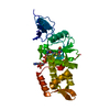
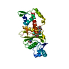
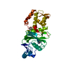
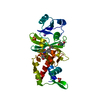
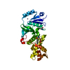
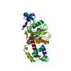
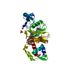
- PDB-1fy7: CRYSTAL STRUCTURE OF YEAST ESA1 HISTONE ACETYLTRANSFERASE DOMAIN ... -
+ Open data

- Basic information
| Entry | Database: PDB / ID: 1fy7 | ||||||
|---|---|---|---|---|---|---|---|
| Title | CRYSTAL STRUCTURE OF YEAST ESA1 HISTONE ACETYLTRANSFERASE DOMAIN COMPLEXED WITH COENZYME A | ||||||
 Components Components | ESA1 HISTONE ACETYLTRANSFERASE | ||||||
 Keywords Keywords | TRANSFERASE / HISTONE ACETYLTRANSFERASE / coenzyme A | ||||||
| Function / homology |  Function and homology information Function and homology informationDNA Damage/Telomere Stress Induced Senescence / Sensing of DNA Double Strand Breaks / piccolo histone acetyltransferase complex / peptide 2-hydroxyisobutyryltransferase activity / histone crotonyltransferase activity / positive regulation of triglyceride biosynthetic process / histone H4 acetyltransferase activity / rDNA heterochromatin formation / DNA-templated transcription elongation / Recruitment and ATM-mediated phosphorylation of repair and signaling proteins at DNA double strand breaks ...DNA Damage/Telomere Stress Induced Senescence / Sensing of DNA Double Strand Breaks / piccolo histone acetyltransferase complex / peptide 2-hydroxyisobutyryltransferase activity / histone crotonyltransferase activity / positive regulation of triglyceride biosynthetic process / histone H4 acetyltransferase activity / rDNA heterochromatin formation / DNA-templated transcription elongation / Recruitment and ATM-mediated phosphorylation of repair and signaling proteins at DNA double strand breaks / histone acetyltransferase activity / NuA4 histone acetyltransferase complex / Estrogen-dependent gene expression / positive regulation of macroautophagy / protein-lysine-acetyltransferase activity / histone acetyltransferase / Transferases; Acyltransferases; Transferring groups other than aminoacyl groups / transcription coregulator activity / positive regulation of transcription elongation by RNA polymerase II / nucleosome / regulation of cell cycle / DNA repair / chromatin binding / regulation of transcription by RNA polymerase II / chromatin / DNA-templated transcription / nucleus Similarity search - Function | ||||||
| Biological species |  | ||||||
| Method |  X-RAY DIFFRACTION / Resolution: 2 Å X-RAY DIFFRACTION / Resolution: 2 Å | ||||||
 Authors Authors | Yan, Y. / Barlev, N.A. / Haley, R.H. / Berger, S.L. / Marmorstein, R. | ||||||
 Citation Citation | Journal: Mol.Cell / Year: 2000 Title: Crystal structure of yeast Esa1 suggests a unified mechanism for catalysis and substrate binding by histone acetyltransferases. Authors: Yan, Y. / Barlev, N.A. / Haley, R.H. / Berger, S.L. / Marmorstein, R. | ||||||
| History |
|
- Structure visualization
| Structure viewer | Molecule: MolmilJmol/JSmol |
|---|
- Downloads & links
-Download
| PDBx/mmCIF format | 1fy7.cif.gz | 75.4 KB | Display | PDBx/mmCIF format |
|---|---|---|---|---|
| PDB format | pdb1fy7.ent.gz | 56.5 KB | Display | PDB format |
| PDBx/mmJSON format | 1fy7.json.gz | Tree view | PDBx/mmJSON format | |
| Others |  Other downloads Other downloads |
-Validation report
| Arichive directory | https://data.pdbj.org/pub/pdb/validation_reports/fy/1fy7ftp://data.pdbj.org/pub/pdb/validation_reports/fy/1fy7 | HTTPS FTP |
|---|
-Related structure data
| Similar structure data |
|---|


-Links
 PDBj
PDBj


- Assembly
| Deposited unit | 
| ||||||||||
|---|---|---|---|---|---|---|---|---|---|---|---|
| 1 |
| ||||||||||
| 2 | 
| ||||||||||
| 3 | x 6
| ||||||||||
| Unit cell |
| ||||||||||
| Details | The biological assembly is a monomer of yeast Esa1 HAT domain bound to coenzyme-A. |
-Components
| #1: Protein | Mass: 33372.414 Da / Num. of mol.: 1 / Fragment: ACETYLTRANSFERASE DOMAIN Source method: isolated from a genetically manipulated source Source: (gene. exp.) Plasmid: PRSETA / Production host:  |
|---|---|
| #2: Chemical | ChemComp-NA /   Mass: 22.990 Da / Num. of mol.: 1 / Source method: obtained synthetically / Formula: Na Mass: 22.990 Da / Num. of mol.: 1 / Source method: obtained synthetically / Formula: Na |
| #3: Chemical | ChemComp-COA /   Mass: 767.534 Da / Num. of mol.: 1 / Source method: obtained synthetically / Formula: C21H36N7O16P3S Mass: 767.534 Da / Num. of mol.: 1 / Source method: obtained synthetically / Formula: C21H36N7O16P3S |
| #4: Water | ChemComp-HOH /  Mass: 18.015 Da / Num. of mol.: 178 / Source method: isolated from a natural source / Formula: H2O Mass: 18.015 Da / Num. of mol.: 178 / Source method: isolated from a natural source / Formula: H2O |
-Experimental details
-Experiment
| Experiment | Method: X-RAY DIFFRACTION / Number of used crystals: 1 |
|---|
- Sample preparation
| Crystal | Density Matthews: 3.73 Å3/Da / Density % sol: 67.03 % | |||||||||||||||||||||||||
|---|---|---|---|---|---|---|---|---|---|---|---|---|---|---|---|---|---|---|---|---|---|---|---|---|---|---|
| Crystal grow | Temperature: 293 K / Method: vapor diffusion, hanging drop / pH: 6.5 Details: amonium phosphate, cacodylate,, pH 6.5, VAPOR DIFFUSION, HANGING DROP, temperature 293.0K | |||||||||||||||||||||||||
| Crystal grow | *PLUS Temperature: 20 ℃ | |||||||||||||||||||||||||
| Components of the solutions | *PLUS
|
-Data collection
| Diffraction | Mean temperature: 93 K |
|---|---|
| Diffraction source | Source: ROTATING ANODE / Type: RIGAKU RU300 / Wavelength: 1.5418 |
| Detector | Type: RIGAKU RAXIS IV / Detector: IMAGE PLATE / Date: Mar 26, 2000 |
| Radiation | Protocol: SINGLE WAVELENGTH / Monochromatic (M) / Laue (L): M / Scattering type: x-ray |
| Radiation wavelength | Wavelength: 1.5418 Å / Relative weight: 1 |
| Reflection | Resolution: 2→20 Å / Num. all: 34558 / Num. obs: 34558 / % possible obs: 99.9 % / Observed criterion σ(F): 0 / Observed criterion σ(I): 0 / Redundancy: 35.9 % / Biso Wilson estimate: 29.13 Å2 / Rmerge(I) obs: 0.063 / Net I/σ(I): 48.6 |
| Reflection shell | Resolution: 2→2.07 Å / Redundancy: 8.8 % / Rmerge(I) obs: 0.272 / Num. unique all: 3413 / % possible all: 99.8 |
| Reflection | *PLUS Num. measured all: 1241495 |
| Reflection shell | *PLUS % possible obs: 99.8 % |
- Processing
| Software |
| |||||||||||||||||||||||||
|---|---|---|---|---|---|---|---|---|---|---|---|---|---|---|---|---|---|---|---|---|---|---|---|---|---|---|
| Refinement | Resolution: 2→20 Å / Cross valid method: THROUGHOUT / σ(F): 2 / Stereochemistry target values: CNS Library / Details: maximum likelihood target using amplitudes
| |||||||||||||||||||||||||
| Refinement step | Cycle: LAST / Resolution: 2→20 Å
| |||||||||||||||||||||||||
| Refine LS restraints |
|
