 Movie
Movie Controller

[English] 日本語
 Yorodumi
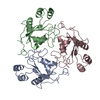
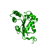
- PDB-1w7w: Structure and mutational analysis of a plant mitochondrial nucleo... -
+ Open data

- Basic information
| Entry | Database: PDB / ID: 1w7w | ||||||
|---|---|---|---|---|---|---|---|
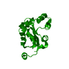
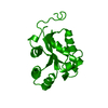
| Title | Structure and mutational analysis of a plant mitochondrial nucleoside diphosphate kinase: identification of residues involved in serine phosphorylation and oligomerization. | ||||||
 Components Components | NUCLEOSIDE DIPHOSPHATE KINASE | ||||||
 Keywords Keywords | TRANSFERASE / MITOCHONDRIAL NUCLEOSIDE DIPHOSPHATE KINASE / NDPK3 / PISUM SATIVUM / KINASE | ||||||
| Function / homology |  Function and homology information Function and homology informationnucleoside-diphosphate kinase / CTP biosynthetic process / UTP biosynthetic process / GTP biosynthetic process / nucleoside diphosphate kinase activity / ATP binding Similarity search - Function | ||||||
| Biological species |   PISUM SATIVUM (garden pea) PISUM SATIVUM (garden pea) | ||||||
| Method |  X-RAY DIFFRACTION / SYNCHROTRON / MOLECULAR REPLACEMENT / Resolution: 2.8 Å X-RAY DIFFRACTION / SYNCHROTRON / MOLECULAR REPLACEMENT / Resolution: 2.8 Å | ||||||
 Authors Authors | Johansson, M. / MacKenzie-Hose, A. / Andersson, I. / Knorpp, C. | ||||||
 Citation Citation | Journal: Plant Physiol. / Year: 2004 Title: Structure and Mutational Analysis of a Plant Mitochondrial Nucleoside Diphosphate Kinase: Identification of Residues Involved in Serine Phosphorylation and Oligomerisation Authors: Johansson, M. / Mackenzie-Hose, A. / Andersson, I. / Knorpp, C. | ||||||
| History |
| ||||||
| Remark 650 | HELIX DETERMINATION METHOD: AUTHOR PROVIDED. |
- Structure visualization
| Structure viewer | Molecule: MolmilJmol/JSmol |
|---|
- Downloads & links
-Download
| PDBx/mmCIF format | 1w7w.cif.gz | 182.1 KB | Display | PDBx/mmCIF format |
|---|---|---|---|---|
| PDB format | pdb1w7w.ent.gz | 146.5 KB | Display | PDB format |
| PDBx/mmJSON format | 1w7w.json.gz | Tree view | PDBx/mmJSON format | |
| Others |  Other downloads Other downloads |
-Validation report
| Summary document | 1w7w_validation.pdf.gz | 476.2 KB | Display | wwPDB validaton report |
|---|---|---|---|---|
| Full document | 1w7w_full_validation.pdf.gz | 496 KB | Display | |
| Data in XML | 1w7w_validation.xml.gz | 34.9 KB | Display | |
| Data in CIF | 1w7w_validation.cif.gz | 46.3 KB | Display | |
| Arichive directory | https://data.pdbj.org/pub/pdb/validation_reports/w7/1w7wftp://data.pdbj.org/pub/pdb/validation_reports/w7/1w7w | HTTPS FTP |
-Related structure data
| Related structure data | 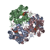 1f6tS S: Starting model for refinement |
|---|---|
| Similar structure data |

-Links
 PDBj
PDBj- Assembly
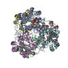
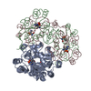
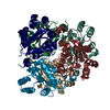
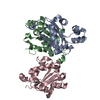
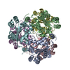
| Deposited unit | 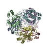
| ||||||||||||
|---|---|---|---|---|---|---|---|---|---|---|---|---|---|
| 1 |
| ||||||||||||
| Unit cell |
| ||||||||||||
| Noncrystallographic symmetry (NCS) | NCS oper:
|
-Components
| #1: Protein | Mass: 20722.404 Da / Num. of mol.: 6 Source method: isolated from a genetically manipulated source Source: (gene. exp.) PISUM SATIVUM (garden pea) / Strain: OREGON SUGAR POD / Plasmid: PPROEX / Production host:  #2: Water | ChemComp-HOH / |  Mass: 18.015 Da / Num. of mol.: 54 / Source method: isolated from a natural source / Formula: H2O Mass: 18.015 Da / Num. of mol.: 54 / Source method: isolated from a natural source / Formula: H2O |
|---|
-Experimental details
-Experiment
| Experiment | Method: X-RAY DIFFRACTION / Number of used crystals: 1 |
|---|
- Sample preparation
| Crystal | Density Matthews: 2 Å3/Da / Density % sol: 39.3 % |
|---|---|
| Crystal grow | pH: 5.5 Details: 16 TO 18% METHYL PENTANEDIOL, 100 MM SODIUM ACETATE, PH 5.5 |
-Data collection
| Diffraction | Mean temperature: 100 K |
|---|---|
| Diffraction source | Source: SYNCHROTRON / Site: ESRF  / Beamline: ID14-4 / Wavelength: 1 / Beamline: ID14-4 / Wavelength: 1 |
| Detector | Type: ADSC CCD / Detector: CCD / Date: Sep 14, 2003 / Details: MIRRORS |
| Radiation | Monochromator: SI(111) / Protocol: SINGLE WAVELENGTH / Monochromatic (M) / Laue (L): M / Scattering type: x-ray |
| Radiation wavelength | Wavelength: 1 Å / Relative weight: 1 |
| Reflection | Resolution: 2.8→20 Å / Num. obs: 26099 / % possible obs: 98.2 % / Observed criterion σ(I): -3 / Redundancy: 5.46 % / Biso Wilson estimate: 50.1 Å2 / Rmerge(I) obs: 0.16 / Net I/σ(I): 9.9 |
| Reflection shell | Resolution: 2.8→2.9 Å / Redundancy: 4.5 % / Rmerge(I) obs: 0.58 / Mean I/σ(I) obs: 1.7 / % possible all: 85.8 |
- Processing
| Software |
| ||||||||||||||||||||||||||||||||||||||||||||||||||||||||||||||||||||||||||||||||
|---|---|---|---|---|---|---|---|---|---|---|---|---|---|---|---|---|---|---|---|---|---|---|---|---|---|---|---|---|---|---|---|---|---|---|---|---|---|---|---|---|---|---|---|---|---|---|---|---|---|---|---|---|---|---|---|---|---|---|---|---|---|---|---|---|---|---|---|---|---|---|---|---|---|---|---|---|---|---|---|---|---|
| Refinement | Method to determine structure: MOLECULAR REPLACEMENT Starting model: PDB ENTRY 1F6T Resolution: 2.8→19.84 Å / Rfactor Rfree error: 0.008 / Data cutoff high absF: 2021609.8 / Isotropic thermal model: RESTRAINED / Cross valid method: THROUGHOUT / σ(F): 0 / Stereochemistry target values: MAXIMUM LIKELIHOOD
| ||||||||||||||||||||||||||||||||||||||||||||||||||||||||||||||||||||||||||||||||
| Solvent computation | Solvent model: FLAT MODEL / Bsol: 20.122 Å2 / ksol: 0.307388 e/Å3 | ||||||||||||||||||||||||||||||||||||||||||||||||||||||||||||||||||||||||||||||||
| Displacement parameters | Biso mean: 39.7 Å2
| ||||||||||||||||||||||||||||||||||||||||||||||||||||||||||||||||||||||||||||||||
| Refine analyze |
| ||||||||||||||||||||||||||||||||||||||||||||||||||||||||||||||||||||||||||||||||
| Refinement step | Cycle: LAST / Resolution: 2.8→19.84 Å
| ||||||||||||||||||||||||||||||||||||||||||||||||||||||||||||||||||||||||||||||||
| Refine LS restraints |
| ||||||||||||||||||||||||||||||||||||||||||||||||||||||||||||||||||||||||||||||||
| LS refinement shell | Resolution: 2.8→2.98 Å / Rfactor Rfree error: 0.032 / Total num. of bins used: 6
| ||||||||||||||||||||||||||||||||||||||||||||||||||||||||||||||||||||||||||||||||
| Xplor file |
|
