 Movie
Movie Controller


+ Open data

- Basic information
| Entry | Database: PDB / ID: 1vz3 | ||||||
|---|---|---|---|---|---|---|---|
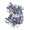
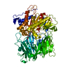
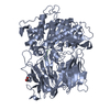
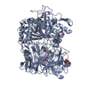
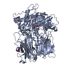
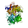
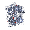
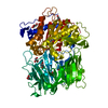
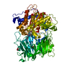
| Title | PROLYL OLIGOPEPTIDASE FROM PORCINE BRAIN, T597C MUTANT | ||||||
 Components Components | PROLYL ENDOPEPTIDASE | ||||||
 Keywords Keywords | HYDROLASE / PROLYL OLIGOPEPTIDASE / AMNESIA / ALPHA/ BETA-HYDROLASE / BETA-PROPELLER / SERINE PROTEASE | ||||||
| Function / homology |  Function and homology information Function and homology informationprolyl oligopeptidase / serine-type endopeptidase activity / proteolysis / cytoplasm Similarity search - Function | ||||||
| Biological species |  | ||||||
| Method |  X-RAY DIFFRACTION / SYNCHROTRON / MOLECULAR REPLACEMENT / Resolution: 1.6 Å X-RAY DIFFRACTION / SYNCHROTRON / MOLECULAR REPLACEMENT / Resolution: 1.6 Å | ||||||
 Authors Authors | Rea, D. / Fulop, V. | ||||||
 Citation Citation | Journal: J.Mol.Biol. / Year: 2004 Title: Concerted Structural Changes in the Peptidase and the Propeller Domains of Prolyl Oligopeptidase are Required for Substrate Binding Authors: Szeltner, Z. / Rea, D. / Juhasz, T. / Renner, V. / Fulop, V. / Polgar, L. #1: Journal: J.Biol.Chem. / Year: 2003Title: Electrostatic Environment at the Active Site of Prolyl Oligopeptidase is Highly Influential During Substrate Binding Authors: Szeltner, Z. / Rea, D. / Renner, V. / Juliano, L. / Fulop, V. / Polgar, L. #2: Journal: J.Biol.Chem. / Year: 2002Title: Electrostatic Effects and Binding Determinants in the Catalysis of Prolyl Oligopeptidase: Site Specific Mutagenesis at the Oxyanion Binding Site Authors: Szeltner, Z. / Rea, D. / Renner, V. / Fulop, V. / Polgar, L. #3: Journal: J.Biol.Chem. / Year: 2002Title: Substrate-Dependent Competency of the Catalytic Triad of Prolyl Oligopeptidase Authors: Szeltner, Z. / Rea, D. / Juhasz, T. / Renner, V. / Mucsi, Z. / Orosz, G. / Fulop, V. / Polgar, L. #4: Journal: J.Biol.Chem. / Year: 2001Title: Structures of Prolyl Oligopeptidase Substrate/ Inhibitor Complexes. Use of Inhibitor Binding for Titration of the Catalytic Histidine Residue Authors: Fulop, V. / Szeltner, Z. / Renner, V. / Polgar, L. #5: Journal: Embo Rep. / Year: 2000Title: Catalysis of Serine Oligopeptidases is Controlled by a Gating Filter Mechanism Authors: Fulop, V. / Szeltner, Z. / Polgar, L. #6: Journal: Cell(Cambridge,Mass.) / Year: 1998Title: Prolyl Oligopeptidase: An Unusual Beta-Propeller Domain Regulates Proteolysis Authors: Fulop, V. / Bocskei, Z. / Polgar, L. | ||||||
| History |
| ||||||
| Remark 700 | SHEET THE SHEET STRUCTURE OF THIS MOLECULE IS BIFURCATED. IN ORDER TO REPRESENT THIS FEATURE IN ... SHEET THE SHEET STRUCTURE OF THIS MOLECULE IS BIFURCATED. IN ORDER TO REPRESENT THIS FEATURE IN THE SHEET RECORDS BELOW, TWO SHEETS ARE DEFINED. |
- Structure visualization
| Structure viewer | Molecule: MolmilJmol/JSmol |
|---|
- Downloads & links
-Download
| PDBx/mmCIF format | 1vz3.cif.gz | 180.6 KB | Display | PDBx/mmCIF format |
|---|---|---|---|---|
| PDB format | pdb1vz3.ent.gz | 141.3 KB | Display | PDB format |
| PDBx/mmJSON format | 1vz3.json.gz | Tree view | PDBx/mmJSON format | |
| Others |  Other downloads Other downloads |
-Validation report
| Arichive directory | https://data.pdbj.org/pub/pdb/validation_reports/vz/1vz3ftp://data.pdbj.org/pub/pdb/validation_reports/vz/1vz3 | HTTPS FTP |
|---|
-Related structure data
| Related structure data | 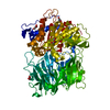 1vz2C  1h2wS S: Starting model for refinement C: citing same article ( |
|---|---|
| Similar structure data |


-Links
 PDBj
PDBj
- Assembly
| Deposited unit | 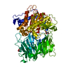
| ||||||||
|---|---|---|---|---|---|---|---|---|---|
| 1 |
| ||||||||
| Unit cell |
|
-Components
| #1: Protein | Mass: 80866.383 Da / Num. of mol.: 1 / Mutation: YES Source method: isolated from a genetically manipulated source Details: ENGINEERED MUTATION THR 597 CYS / Source: (gene. exp.)  | ||||||
|---|---|---|---|---|---|---|---|
| #2: Chemical | ChemComp-GOL /   Mass: 92.094 Da / Num. of mol.: 5 / Source method: obtained synthetically / Formula: C3H8O3 Mass: 92.094 Da / Num. of mol.: 5 / Source method: obtained synthetically / Formula: C3H8O3#3: Water | ChemComp-HOH / |  Mass: 18.015 Da / Num. of mol.: 1161 / Source method: isolated from a natural source / Formula: H2O Mass: 18.015 Da / Num. of mol.: 1161 / Source method: isolated from a natural source / Formula: H2OCompound details | ENGINEERED MUTATION IN CHAIN A, THR 597 CYS IN CHAIN A CLEAVES PEPTIDE BONDS ON THE C-TERMINAL SIDE ...ENGINEERED | Has protein modification | Y | |
-Experimental details
-Experiment
| Experiment | Method: X-RAY DIFFRACTION / Number of used crystals: 1 |
|---|
- Sample preparation
| Crystal | Density Matthews: 2.5 Å3/Da / Density % sol: 44 % |
|---|---|
| Crystal grow | pH: 8.5 / Details: SEE REMARK 1, REFERENCE 6, pH 8.50 |
-Data collection
| Diffraction | Mean temperature: 100 K |
|---|---|
| Diffraction source | Source: SYNCHROTRON / Site: SRS  / Beamline: PX14.1 / Wavelength: 1.488 / Beamline: PX14.1 / Wavelength: 1.488 |
| Detector | Type: ADSC CCD / Detector: CCD / Date: Jul 10, 2003 / Details: MIRRORS |
| Radiation | Protocol: SINGLE WAVELENGTH / Monochromatic (M) / Laue (L): M / Scattering type: x-ray |
| Radiation wavelength | Wavelength: 1.488 Å / Relative weight: 1 |
| Reflection | Resolution: 1.6→26 Å / Num. obs: 105437 / % possible obs: 99 % / Observed criterion σ(I): -3 / Redundancy: 3.7 % / Biso Wilson estimate: 21.9 Å2 / Rmerge(I) obs: 0.06 / Net I/σ(I): 23.3 |
| Reflection shell | Resolution: 1.6→1.66 Å / Redundancy: 2.3 % / Rmerge(I) obs: 0.216 / Mean I/σ(I) obs: 3.2 / % possible all: 99.3 |
- Processing
| Software |
| ||||||||||||||||||||
|---|---|---|---|---|---|---|---|---|---|---|---|---|---|---|---|---|---|---|---|---|---|
| Refinement | Method to determine structure: MOLECULAR REPLACEMENT Starting model: PDB ENTRY 1H2W Resolution: 1.6→26 Å / SU B: 2.7 / SU ML: 0.05 / Cross valid method: THROUGHOUT / σ(F): 0 / ESU R: 0.08 / ESU R Free: 0.08
| ||||||||||||||||||||
| Displacement parameters | Biso mean: 21.5 Å2
| ||||||||||||||||||||
| Refinement step | Cycle: LAST / Resolution: 1.6→26 Å
|
