 Movie
Movie Controller

[English] 日本語
 Yorodumi
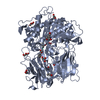
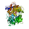
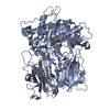
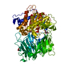
- PDB-1e8m: PROLYL OLIGOPEPTIDASE FROM PORCINE BRAIN, MUTANT, COMPLEXED WITH ... -
+ Open data

- Basic information
| Entry | Database: PDB / ID: 1e8m | ||||||
|---|---|---|---|---|---|---|---|
| Title | PROLYL OLIGOPEPTIDASE FROM PORCINE BRAIN, MUTANT, COMPLEXED WITH INHIBITOR | ||||||
 Components Components | PROLYL ENDOPEPTIDASE | ||||||
 Keywords Keywords | HYDROLASE/HYDROLASE INHIBITOR / HYDROLASE / PROLYL OLIGOPEPTIDASE / AMNESIA / ALPHA/ BETA-HYDROLASE / BETA-PROPELLER / HYDROLASE-HYDROLASE INHIBITOR COMPLEX | ||||||
| Function / homology |  Function and homology information Function and homology informationprolyl oligopeptidase / serine-type endopeptidase activity / proteolysis / cytoplasm Similarity search - Function | ||||||
| Biological species |  | ||||||
| Method |  X-RAY DIFFRACTION / SYNCHROTRON / MOLECULAR REPLACEMENT / Resolution: 1.5 Å X-RAY DIFFRACTION / SYNCHROTRON / MOLECULAR REPLACEMENT / Resolution: 1.5 Å | ||||||
 Authors Authors | Fulop, V. | ||||||
 Citation Citation | Journal: J.Biol.Chem. / Year: 2001 Title: Structures of Prolyl Oligopeptidase Substrate/ Inhibitor Complexes. Use of Inhibitor Binding for Titration of the Catalytic Histidine Residue Authors: Fulop, V. / Szeltner, Z. / Renner, V. / Polgar, L. #1: Journal: Embo Rep. / Year: 2000Title: Catalysis of Serine Oligopeptidases is Controlled by a Gating Filter Mechanism Authors: Fulop, V. / Szeltner, Z. / Polgar, L. #2: Journal: Cell(Cambridge,Mass.) / Year: 1998Title: Prolyl Oligopeptidase: An Unusual Beta-Propeller Domain Regulates Proteolysis Authors: Fulop, V. / Bocskei, Z. / Polgar, L. | ||||||
| History |
|
- Structure visualization
| Structure viewer | Molecule: MolmilJmol/JSmol |
|---|
- Downloads & links
-Download
| PDBx/mmCIF format | 1e8m.cif.gz | 181.6 KB | Display | PDBx/mmCIF format |
|---|---|---|---|---|
| PDB format | pdb1e8m.ent.gz | 141.2 KB | Display | PDB format |
| PDBx/mmJSON format | 1e8m.json.gz | Tree view | PDBx/mmJSON format | |
| Others |  Other downloads Other downloads |
-Validation report
| Arichive directory | https://data.pdbj.org/pub/pdb/validation_reports/e8/1e8mftp://data.pdbj.org/pub/pdb/validation_reports/e8/1e8m | HTTPS FTP |
|---|
-Related structure data
| Related structure data | 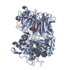 1e8nC  1qfmS C: citing same article ( S: Starting model for refinement |
|---|---|
| Similar structure data |


-Links
 PDBj
PDBj
- Assembly
| Deposited unit | 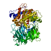
| ||||||||
|---|---|---|---|---|---|---|---|---|---|
| 1 |
| ||||||||
| Unit cell |
|
-Components
| #1: Protein | Mass: 80848.344 Da / Num. of mol.: 1 / Mutation: YES Source method: isolated from a genetically manipulated source Source: (gene. exp.)  |
|---|---|
| #2: Chemical | ChemComp-GOL /   Mass: 92.094 Da / Num. of mol.: 1 / Source method: obtained synthetically / Formula: C3H8O3 Mass: 92.094 Da / Num. of mol.: 1 / Source method: obtained synthetically / Formula: C3H8O3 |
| #3: Chemical | ChemComp-P0H / 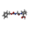  Type: peptide-like, Peptide-like / Class: Inhibitor / Mass: 306.314 Da / Num. of mol.: 1 / Source method: obtained synthetically / Formula: C15H18N2O5 / References: N-[(benzyloxy)carbonyl]glycyl-L-proline Type: peptide-like, Peptide-like / Class: Inhibitor / Mass: 306.314 Da / Num. of mol.: 1 / Source method: obtained synthetically / Formula: C15H18N2O5 / References: N-[(benzyloxy)carbonyl]glycyl-L-proline |
| #4: Water | ChemComp-HOH /  Mass: 18.015 Da / Num. of mol.: 1203 / Source method: isolated from a natural source / Formula: H2O Mass: 18.015 Da / Num. of mol.: 1203 / Source method: isolated from a natural source / Formula: H2O |
| Compound details | CHAIN A CONTAINS ENGINEERED |
-Experimental details
-Experiment
| Experiment | Method: X-RAY DIFFRACTION / Number of used crystals: 1 |
|---|
- Sample preparation
| Crystal | Density Matthews: 2.4 Å3/Da / Density % sol: 48 % | ||||||||||||||||||||||||||||||||||||||||||
|---|---|---|---|---|---|---|---|---|---|---|---|---|---|---|---|---|---|---|---|---|---|---|---|---|---|---|---|---|---|---|---|---|---|---|---|---|---|---|---|---|---|---|---|
| Crystal grow | pH: 8.5 / Details: SEE REFERENCE, PH 8.50 | ||||||||||||||||||||||||||||||||||||||||||
| Crystal grow | *PLUS Temperature: 4 ℃ / Method: vapor diffusion, hanging drop / Details: Fulop, V., (1998) Cell(Cambridge,Mass.), 94, 161. | ||||||||||||||||||||||||||||||||||||||||||
| Components of the solutions | *PLUS
|
-Data collection
| Diffraction | Mean temperature: 100 K |
|---|---|
| Diffraction source | Source: SYNCHROTRON / Site: EMBL/DESY, HAMBURG  / Beamline: X11 / Wavelength: 0.909 / Beamline: X11 / Wavelength: 0.909 |
| Detector | Type: MARRESEARCH / Detector: IMAGE PLATE / Date: Jul 15, 1999 / Details: MIRRORS |
| Radiation | Protocol: SINGLE WAVELENGTH / Monochromatic (M) / Laue (L): M / Scattering type: x-ray |
| Radiation wavelength | Wavelength: 0.909 Å / Relative weight: 1 |
| Reflection | Resolution: 1.5→18 Å / Num. obs: 124438 / % possible obs: 95.4 % / Observed criterion σ(I): -3 / Redundancy: 3.8 % / Biso Wilson estimate: 13.7 Å2 / Rsym value: 0.032 / Net I/σ(I): 36 |
| Reflection shell | Resolution: 1.5→1.54 Å / Redundancy: 3.6 % / Mean I/σ(I) obs: 8 / Rsym value: 0.128 / % possible all: 90.7 |
| Reflection | *PLUS Num. measured all: 470813 / Rmerge(I) obs: 0.032 |
- Processing
| Software |
| ||||||||||||||||||||||||||||||||||||||||||||||||||||||||||||
|---|---|---|---|---|---|---|---|---|---|---|---|---|---|---|---|---|---|---|---|---|---|---|---|---|---|---|---|---|---|---|---|---|---|---|---|---|---|---|---|---|---|---|---|---|---|---|---|---|---|---|---|---|---|---|---|---|---|---|---|---|---|
| Refinement | Method to determine structure: MOLECULAR REPLACEMENT Starting model: PDB ENTRY 1QFM Resolution: 1.5→18 Å / Cross valid method: THROUGHOUT / σ(F): 2
| ||||||||||||||||||||||||||||||||||||||||||||||||||||||||||||
| Displacement parameters | Biso mean: 14.5 Å2 | ||||||||||||||||||||||||||||||||||||||||||||||||||||||||||||
| Refinement step | Cycle: LAST / Resolution: 1.5→18 Å
| ||||||||||||||||||||||||||||||||||||||||||||||||||||||||||||
| Refine LS restraints |
| ||||||||||||||||||||||||||||||||||||||||||||||||||||||||||||
| Software | *PLUS Name: X-PLOR / Version: 3.851 / Classification: refinement | ||||||||||||||||||||||||||||||||||||||||||||||||||||||||||||
| Refinement | *PLUS Rfactor all: 0.195 / Rfactor obs: 0.193 / Rfactor Rfree: 0.217 / Num. reflection all: 124438 | ||||||||||||||||||||||||||||||||||||||||||||||||||||||||||||
| Solvent computation | *PLUS | ||||||||||||||||||||||||||||||||||||||||||||||||||||||||||||
| Displacement parameters | *PLUS | ||||||||||||||||||||||||||||||||||||||||||||||||||||||||||||
| Refine LS restraints | *PLUS
|
