 Movie
Movie Controller


+ Open data

- Basic information
| Entry | Database: PDB / ID: 1qfm | ||||||
|---|---|---|---|---|---|---|---|
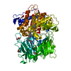
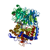
| Title | PROLYL OLIGOPEPTIDASE FROM PORCINE MUSCLE | ||||||
 Components Components | PROTEIN (PROLYL OLIGOPEPTIDASE) | ||||||
 Keywords Keywords | HYDROLASE / PROLYL OLIGOPEPTIDASE / AMNESIA / ALPHA/BETA-HYDROLASE / BETA-PROPELLER | ||||||
| Function / homology |  Function and homology information Function and homology informationprolyl oligopeptidase / serine-type endopeptidase activity / proteolysis / cytoplasm Similarity search - Function | ||||||
| Biological species |  | ||||||
| Method |  X-RAY DIFFRACTION / SYNCHROTRON / MIR / Resolution: 1.4 Å X-RAY DIFFRACTION / SYNCHROTRON / MIR / Resolution: 1.4 Å | ||||||
 Authors Authors | Fulop, V. | ||||||
 Citation Citation | Journal: Cell(Cambridge,Mass.) / Year: 1998 Title: Prolyl oligopeptidase: an unusual beta-propeller domain regulates proteolysis. Authors: Fulop, V. / Bocskei, Z. / Polgar, L. | ||||||
| History |
|
- Structure visualization
| Structure viewer | Molecule: MolmilJmol/JSmol |
|---|
- Downloads & links
-Download
| PDBx/mmCIF format | 1qfm.cif.gz | 172.1 KB | Display | PDBx/mmCIF format |
|---|---|---|---|---|
| PDB format | pdb1qfm.ent.gz | 135.9 KB | Display | PDB format |
| PDBx/mmJSON format | 1qfm.json.gz | Tree view | PDBx/mmJSON format | |
| Others |  Other downloads Other downloads |
-Validation report
| Arichive directory | https://data.pdbj.org/pub/pdb/validation_reports/qf/1qfmftp://data.pdbj.org/pub/pdb/validation_reports/qf/1qfm | HTTPS FTP |
|---|
-Related structure data


-Links
 PDBj
PDBj
- Assembly
| Deposited unit | 
| ||||||||
|---|---|---|---|---|---|---|---|---|---|
| 1 |
| ||||||||
| Unit cell |
|
-Components
| #1: Protein | Mass: 80976.344 Da / Num. of mol.: 1 / Source method: isolated from a natural source Details: PORCINE BRAIN SEQUENCE WAS USED FOR STRUCTURE DETERMINATION AND REFINEMENT Source: (natural) | ||||||||||
|---|---|---|---|---|---|---|---|---|---|---|---|
| #2: Chemical |   Mass: 124.159 Da / Num. of mol.: 2 / Source method: obtained synthetically / Formula: C3H8O3S Mass: 124.159 Da / Num. of mol.: 2 / Source method: obtained synthetically / Formula: C3H8O3S#3: Chemical |   Mass: 108.159 Da / Num. of mol.: 2 / Source method: obtained synthetically / Formula: C3H8O2S Mass: 108.159 Da / Num. of mol.: 2 / Source method: obtained synthetically / Formula: C3H8O2S#4: Chemical | ChemComp-GOL /   Mass: 92.094 Da / Num. of mol.: 4 / Source method: obtained synthetically / Formula: C3H8O3 Mass: 92.094 Da / Num. of mol.: 4 / Source method: obtained synthetically / Formula: C3H8O3#5: Water | ChemComp-HOH / |  Mass: 18.015 Da / Num. of mol.: 887 / Source method: isolated from a natural source / Formula: H2O Mass: 18.015 Da / Num. of mol.: 887 / Source method: isolated from a natural source / Formula: H2ONonpolymer details | 1-THIOXY-GLYCEROL (SGL781) IS COVALENTLY BOUND TO SER554 MONOTHIOGLYCEROL IS COVALENTLY BOUND TO ...1-THIOXY-GLYCEROL (SGL781) IS COVALENTLY | Sequence details | CYS25, CYS57 AND CYS255 ARE OXIDIZED WITH ONE OXYGEN ATOM. CYS175 AND CYS601 ARE OXIDIZED WITH TWO ...CYS25, CYS57 AND CYS255 ARE OXIDIZED WITH ONE OXYGEN ATOM. CYS175 AND CYS601 ARE OXIDIZED WITH TWO OXYGEN ATOMS. | |
-Experimental details
-Experiment
| Experiment | Method: X-RAY DIFFRACTION / Number of used crystals: 1 |
|---|
- Sample preparation
| Crystal | Density Matthews: 2.4 Å3/Da / Density % sol: 48 % | ||||||||||||||||||||||||||||||||||||||||||
|---|---|---|---|---|---|---|---|---|---|---|---|---|---|---|---|---|---|---|---|---|---|---|---|---|---|---|---|---|---|---|---|---|---|---|---|---|---|---|---|---|---|---|---|
| Crystal grow | pH: 8.5 / Details: SEE REFERENCE, pH 8.50 | ||||||||||||||||||||||||||||||||||||||||||
| Crystal | *PLUS Density % sol: 48 % | ||||||||||||||||||||||||||||||||||||||||||
| Crystal grow | *PLUS Temperature: 4 ℃ / pH: 8.5 / Method: vapor diffusion, hanging drop | ||||||||||||||||||||||||||||||||||||||||||
| Components of the solutions | *PLUS
|
-Data collection
| Diffraction | Mean temperature: 100 K |
|---|---|
| Diffraction source | Source: SYNCHROTRON / Site: SRS  / Beamline: PX9.6 / Wavelength: 0.87 / Beamline: PX9.6 / Wavelength: 0.87 |
| Detector | Type: MARRESEARCH / Detector: IMAGE PLATE / Date: Jun 1, 1997 / Details: MIRRORS |
| Radiation | Protocol: SINGLE WAVELENGTH / Monochromatic (M) / Laue (L): M / Scattering type: x-ray |
| Radiation wavelength | Wavelength: 0.87 Å / Relative weight: 1 |
| Reflection | Resolution: 1.4→30 Å / Num. obs: 140350 / % possible obs: 91 % / Observed criterion σ(I): -3 / Redundancy: 2.6 % / Biso Wilson estimate: 14.7 Å2 / Rmerge(I) obs: 0.057 / Rsym value: 0.057 / Net I/σ(I): 16.4 |
| Reflection shell | Resolution: 1.4→1.44 Å / Redundancy: 2.4 % / Rmerge(I) obs: 0.324 / Mean I/σ(I) obs: 2.4 / Rsym value: 0.324 / % possible all: 80 |
| Reflection | *PLUS Highest resolution: 1.4 Å / Lowest resolution: 30 Å / % possible obs: 91 % / Num. measured all: 366752 |
- Processing
| Software |
| ||||||||||||||||||||||||||||||||||||||||||||||||||||||||||||
|---|---|---|---|---|---|---|---|---|---|---|---|---|---|---|---|---|---|---|---|---|---|---|---|---|---|---|---|---|---|---|---|---|---|---|---|---|---|---|---|---|---|---|---|---|---|---|---|---|---|---|---|---|---|---|---|---|---|---|---|---|---|
| Refinement | Method to determine structure: MIR / Resolution: 1.4→30 Å / Cross valid method: THROUGHOUT / σ(F): 2
| ||||||||||||||||||||||||||||||||||||||||||||||||||||||||||||
| Displacement parameters | Biso mean: 13.5 Å2 | ||||||||||||||||||||||||||||||||||||||||||||||||||||||||||||
| Refinement step | Cycle: LAST / Resolution: 1.4→30 Å
| ||||||||||||||||||||||||||||||||||||||||||||||||||||||||||||
| Refine LS restraints |
| ||||||||||||||||||||||||||||||||||||||||||||||||||||||||||||
| Software | *PLUS Name: X-PLOR / Version: 3.851 / Classification: refinement | ||||||||||||||||||||||||||||||||||||||||||||||||||||||||||||
| Refinement | *PLUS Highest resolution: 1.4 Å / Lowest resolution: 30 Å / Rfactor obs: 0.19 / Rfactor Rwork: 0.19 | ||||||||||||||||||||||||||||||||||||||||||||||||||||||||||||
| Solvent computation | *PLUS | ||||||||||||||||||||||||||||||||||||||||||||||||||||||||||||
| Displacement parameters | *PLUS |
